3HN8

Crystal structure of synaptotagmin


Experimental Data Snapshot

  • Method: X-RAY DIFFRACTION
  • Resolution: 3.50 Å
  • R-Value Free: 0.259 
  • R-Value Work: 0.238 
  • R-Value Observed: 0.240 

wwPDB Validation   3D Report Full Report


This is version 1.3 of the entry. See complete history


Literature

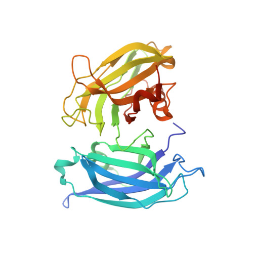
Molecular mechanism of the synaptotagmin-SNARE interaction in Ca2+-triggered vesicle fusion.

Vrljic, M.Strop, P.Ernst, J.A.Sutton, R.B.Chu, S.Brunger, A.T.

(2010) Nat Struct Mol Biol 17: 325-331

  • DOI: https://doi.org/10.1038/nsmb.1764
  • Primary Citation of Related Structures:  
    3HN8

  • PubMed Abstract: 

    In neurons, SNAREs, synaptotagmin and other factors catalyze Ca(2+)-triggered fusion of vesicles with the plasma membrane. The molecular mechanism of this process, especially the interaction between synaptotagmin and SNAREs, remains an enigma. Here we characterized this interaction by single-molecule fluorescence microscopy and crystallography. The two rigid Ca(2+)-binding domains of synaptotagmin 3 (Syt3) undergo large relative motions in solution. Interaction with SNARE complex amplifies a particular state of the two domains that is further enhanced by Ca(2+). This state is represented by the first SNARE-induced Ca(2+)-bound crystal structure of a synaptotagmin fragment containing both domains. The arrangement of the Ca(2+)-binding loops of this structure of Syt3 matches that of SNARE-bound Syt1, suggesting a conserved feature of synaptotagmins. The loops resemble the membrane-interacting loops of certain viral fusion proteins in the postfusion state, suggesting unexpected similarities between both fusion systems.


  • Organizational Affiliation

    Department of Molecular and Cellular Physiology, Stanford University, Stanford, California, USA.


Macromolecules
Find similar proteins by:  (by identity cutoff)  |  3D Structure
Entity ID: 1
MoleculeChains Sequence LengthOrganismDetailsImage
Synaptotagmin-3
A, B, C
296Rattus norvegicusMutation(s): 0 
Gene Names: Syt3
Membrane Entity: Yes 
UniProt
Find proteins for P40748 (Rattus norvegicus)
Explore P40748 
Go to UniProtKB:  P40748
Entity Groups  
Sequence Clusters30% Identity50% Identity70% Identity90% Identity95% Identity100% Identity
UniProt GroupP40748
Sequence Annotations
Expand
  • Reference Sequence
Small Molecules
Ligands 2 Unique
IDChains Name / Formula / InChI Key2D Diagram3D Interactions
ZN
Query on ZN

Download Ideal Coordinates CCD File 
J [auth A],
Q [auth B],
X [auth C]
ZINC ION
Zn
PTFCDOFLOPIGGS-UHFFFAOYSA-N
CA
Query on CA

Download Ideal Coordinates CCD File 
D [auth A]
E [auth A]
F [auth A]
G [auth A]
H [auth A]
D [auth A],
E [auth A],
F [auth A],
G [auth A],
H [auth A],
I [auth A],
K [auth B],
L [auth B],
M [auth B],
N [auth B],
O [auth B],
P [auth B],
R [auth C],
S [auth C],
T [auth C],
U [auth C],
V [auth C],
W [auth C]
CALCIUM ION
Ca
BHPQYMZQTOCNFJ-UHFFFAOYSA-N
Experimental Data & Validation

Experimental Data

  • Method: X-RAY DIFFRACTION
  • Resolution: 3.50 Å
  • R-Value Free: 0.259 
  • R-Value Work: 0.238 
  • R-Value Observed: 0.240 
  • Space Group: P 41 21 2
Unit Cell:
Length ( Å )Angle ( ˚ )
a = 205.675α = 90
b = 205.675β = 90
c = 143.125γ = 90
Software Package:
Software NamePurpose
DENZOdata reduction
SCALEPACKdata scaling
PHENIXrefinement
PDB_EXTRACTdata extraction
HKL-2000data collection
PHASERphasing

Structure Validation

View Full Validation Report


Entry History 

Deposition Data

Revision History  (Full details and data files)

  • Version 1.0: 2010-02-23
    Type: Initial release
  • Version 1.1: 2011-07-13
    Changes: Version format compliance
  • Version 1.2: 2012-03-21
    Changes: Database references
  • Version 1.3: 2017-11-01
    Changes: Refinement description