3HGP | pdb_00003hgp

Structure of porcine pancreatic elastase complexed with a potent peptidyl inhibitor FR130180 determined by high resolution crystallography


Experimental Data Snapshot

  • Method: X-RAY DIFFRACTION
  • Resolution: 0.94 Å
  • R-Value Free: 
    0.128 (Depositor), 0.121 (DCC) 
  • R-Value Work: 
    0.107 (Depositor), 0.101 (DCC) 

Starting Model: experimental
View more details

wwPDB Validation 3D Report Full Report

Validation slider image for 3HGP

Ligand Structure Quality Assessment 


This is version 1.3 of the entry. See complete history

Literature

Combined High-Resolution Neutron and X-ray Analysis of Inhibited Elastase Confirms the Active-Site Oxyanion Hole but Rules against a Low-Barrier Hydrogen Bond

Tamada, T.Kinoshita, T.Kurihara, K.Adachi, M.Ohhara, T.Imai, K.Kuroki, R.Tada, T.

(2009) J Am Chem Soc 131: 11033-11040

  • DOI: https://doi.org/10.1021/ja9028846
  • Primary Citation Related Structures: 
    3HGN, 3HGP

  • PubMed Abstract: 

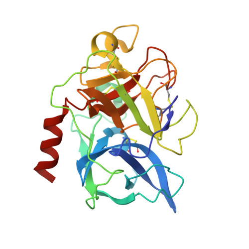
    To help resolve long-standing questions regarding the catalytic activity of the serine proteases, the structure of porcine pancreatic elastase has been analyzed by high-resolution neutron and X-ray crystallography. To mimic the tetrahedral transition intermediate, a peptidic inhibitor was used. A single large crystal was used to collect room-temperature neutron data to 1.65 A resolution and X-ray data to 1.20 A resolution. Another crystal provided a low-temperature X-ray data set to 0.94 A resolution. The neutron data are to higher resolution than previously reported for a serine protease and the X-ray data are comparable with other studies. The neutron and X-ray data show that the hydrogen bond between His57 and Asp102 (chymotrypsin numbering) is 2.60 A in length and that the hydrogen-bonding hydrogen is 0.80-0.96 A from the histidine nitrogen. This is not consistent with a low-barrier hydrogen which is predicted to have the hydrogen midway between the donor and acceptor atom. The observed interaction between His57 and Asp102 is essentially a short but conventional hydrogen bond, sometimes described as a short ionic hydrogen bond. The neutron analysis also shows that the oxygen of the oxopropyl group of the inhibitor is present as an oxygen anion rather than a hydroxyl group, supporting the role of the "oxyanion hole" in stabilizing the tetrahedral intermediate in catalysis.


  • Organizational Affiliation
    • Quantum Beam Science Directorate, Japan Atomic Energy Agency, 2-4 Shirakata-Shirane, Tokai, Ibaraki 319-1195, Japan.

Macromolecule Content 

  • Total Structure Weight: 26.58 kDa 
  • Atom Count: 2,290 
  • Modeled Residue Count: 240 
  • Deposited Residue Count: 240 
  • Unique protein chains: 1

Macromolecules

Find similar proteins by:|  3D Structure
Entity ID: 1
MoleculeChains  Sequence LengthOrganismDetailsImage
Elastase-1240Sus scrofaMutation(s): 0 
EC: 3.4.21.36
UniProt
Find proteins for P00772 (Sus scrofa)
Explore P00772 
Go to UniProtKB:  P00772
Entity Groups
Sequence Clusters30% Identity50% Identity70% Identity90% Identity95% Identity100% Identity
UniProt GroupP00772
Sequence Annotations
Expand
Reference Sequence

Small Molecules

Ligands 3 Unique
IDChains Name / Formula / InChI Key2D Diagram3D Interactions
FRW

Query on FRW


Download:Ideal Coordinates CCD File
B [auth A]4-[[(2S)-3-methyl-1-oxo-1-[(2S)-2-[[(3S)-1,1,1-trifluoro-4-methyl-2-oxo-pentan-3-yl]carbamoyl]pyrrolidin-1-yl]butan-2-yl]carbamoyl]benzoic acid
C24 H30 F3 N3 O6
FGNPKLCZJAUKFU-BZSNNMDCSA-N
SO4

Query on SO4


Download:Ideal Coordinates CCD File
D [auth A]SULFATE ION
O4 S
QAOWNCQODCNURD-UHFFFAOYSA-L
CA

Query on CA


Download:Ideal Coordinates CCD File
C [auth A]CALCIUM ION
Ca
BHPQYMZQTOCNFJ-UHFFFAOYSA-N

Experimental Data & Validation

Experimental Data

  • Method: X-RAY DIFFRACTION
  • Resolution: 0.94 Å
  • R-Value Free:  0.128 (Depositor), 0.121 (DCC) 
  • R-Value Work:  0.107 (Depositor), 0.101 (DCC) 
Space Group: P 21 21 21
Unit Cell:
Length ( Å )Angle ( ˚ )
a = 49.859α = 90
b = 57.049β = 90
c = 74.333γ = 90
Software Package:
Software NamePurpose
ADSCdata collection
AMoREphasing
SHELXL-97refinement
HKL-2000data reduction
HKL-2000data scaling

Structure Validation

View Full Validation Report


Ligand Structure Quality Assessment 


Entry History 

Deposition Data

Revision History  (Full details and data files)

  • Version 1.0: 2009-07-28
    Type: Initial release
  • Version 1.1: 2011-07-13
    Changes: Version format compliance
  • Version 1.2: 2023-11-01
    Changes: Data collection, Database references, Derived calculations, Refinement description
  • Version 1.3: 2024-10-09
    Changes: Structure summary