


Crystal Structure of human ADP-ribosylhydrolase 1
Koch-Nolte, F., Mueller-Dieckmann, J., Weiss, M.S., Mueller-Dieckmann, C.To be published.
Experimental Data Snapshot
Entity ID: 1 | |||||
|---|---|---|---|---|---|
| Molecule | Chains | Sequence Length | Organism | Details | Image |
| Protein ADP-ribosylarginine hydrolase | 357 | Homo sapiens | Mutation(s): 0 EC: 3.2.2.19 | 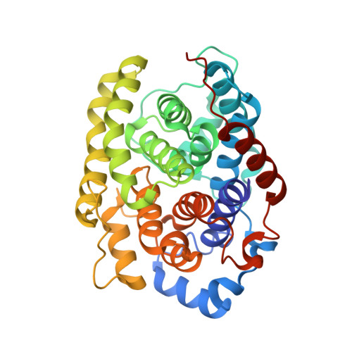 | |
UniProt & NIH Common Fund Data Resources | |||||
PHAROS: P54922 GTEx: ENSG00000144843 | |||||
Entity Groups | |||||
| Sequence Clusters | 30% Identity50% Identity70% Identity90% Identity95% Identity100% Identity | ||||
| UniProt Group | P54922 | ||||
Sequence AnnotationsExpand | |||||
Reference Sequence | |||||
| Ligands 4 Unique | |||||
|---|---|---|---|---|---|
| ID | Chains | Name / Formula / InChI Key | 2D Diagram | 3D Interactions | |
| ADP Download:Ideal Coordinates CCD File | B [auth A] | ADENOSINE-5'-DIPHOSPHATE C10 H15 N5 O10 P2 XTWYTFMLZFPYCI-KQYNXXCUSA-N |  | ||
| K Download:Ideal Coordinates CCD File | C [auth A] | POTASSIUM ION K NPYPAHLBTDXSSS-UHFFFAOYSA-N |  | ||
| CL Download:Ideal Coordinates CCD File | D [auth A] | CHLORIDE ION Cl VEXZGXHMUGYJMC-UHFFFAOYSA-M |  | ||
| MG Download:Ideal Coordinates CCD File | E [auth A] | MAGNESIUM ION Mg JLVVSXFLKOJNIY-UHFFFAOYSA-N |  | ||
| Length ( Å ) | Angle ( ˚ ) |
|---|---|
| a = 45.28 | α = 90 |
| b = 52.79 | β = 90 |
| c = 140.34 | γ = 90 |
| Software Name | Purpose |
|---|---|
| REFMAC | refinement |


