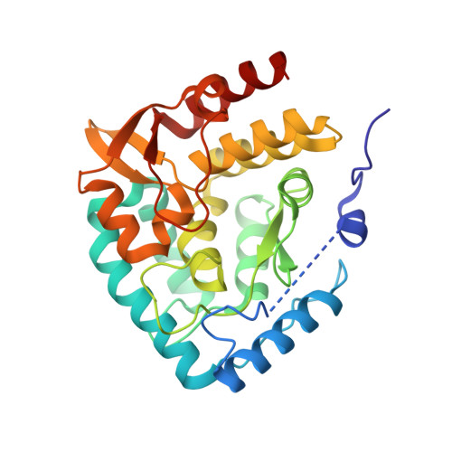
3HF6

Crystal structure of human tryptophan hydroxylase type 1 with bound LP-521834 and FE


Experimental Data Snapshot

  • Method: X-RAY DIFFRACTION
  • Resolution: 1.80 Å
  • R-Value Free: 0.217 
  • R-Value Work: 0.181 

wwPDB Validation   3D Report Full Report


Ligand Structure Quality Assessment 


This is version 1.2 of the entry. See complete history


Literature

Mechanism of Inhibition of Novel Tryptophan Hydroxylase Inhibitors Revealed by Co-crystal Structures and Kinetic Analysis

Cianchetta, G.Stouch, T.Yu, W.Shi, Z.-C.Tari, L.W.Swanson, R.V.Hunter, M.J.Hoffman, I.D.Liu, Q.

(2010) Bioorg Med Chem Lett 4: 19-26

  • DOI: https://doi.org/10.1016/j.bmcl.2009.07.005
  • Primary Citation of Related Structures:  
    3HF6

  • PubMed Abstract: 

    Tryptophan hydroxylase (TPH) is a key enzyme in the synthesis of serotonin. As a neurotransmitter, serotonin plays important physiological roles both peripherally and centrally. Here we describe the discovery of substituted triazines as a novel class of tryptophan hydroxylase inhibitors. This class of TPH inhibitors can selectively reduce serotonin levels in murine intestine after oral administration without affecting levels in the brain. These TPH inhibitors may provide novel treatments for gastrointestinal disorders associated with dysregulation of the serotonergic system, such as chemotherapy-induced emesis and irritable bowel syndrome.


  • Organizational Affiliation

    Lexicon Pharmaceuticals, 350 Carter Road, Princeton, NJ 08540, USA. hjin@lexpharma.com


Macromolecules
Find similar proteins by:  (by identity cutoff)  |  3D Structure
Entity ID: 1
MoleculeChains Sequence LengthOrganismDetailsImage
Tryptophan 5-hydroxylase 1290Homo sapiensMutation(s): 0 
EC: 1.14.16.4
UniProt & NIH Common Fund Data Resources
Find proteins for P17752 (Homo sapiens)
Explore P17752 
Go to UniProtKB:  P17752
PHAROS:  P17752
GTEx:  ENSG00000129167 
Entity Groups  
Sequence Clusters30% Identity50% Identity70% Identity90% Identity95% Identity100% Identity
UniProt GroupP17752
Sequence Annotations
Expand
  • Reference Sequence
Small Molecules
Ligands 2 Unique
IDChains Name / Formula / InChI Key2D Diagram3D Interactions
LX0
Query on LX0

Download Ideal Coordinates CCD File 
C [auth A]4-(4-amino-6-{[(1R)-1-naphthalen-2-ylethyl]amino}-1,3,5-triazin-2-yl)-L-phenylalanine
C24 H24 N6 O2
UXYOCRLZKBFANG-VLIAUNLRSA-N
FE
Query on FE

Download Ideal Coordinates CCD File 
B [auth A]FE (III) ION
Fe
VTLYFUHAOXGGBS-UHFFFAOYSA-N
Binding Affinity Annotations 
IDSourceBinding Affinity
LX0 BindingDB:  3HF6 IC50: 26 (nM) from 1 assay(s)
PDBBind:  3HF6 IC50: 26 (nM) from 1 assay(s)
Binding MOAD:  3HF6 IC50: 26 (nM) from 1 assay(s)
Experimental Data & Validation

Experimental Data

  • Method: X-RAY DIFFRACTION
  • Resolution: 1.80 Å
  • R-Value Free: 0.217 
  • R-Value Work: 0.181 
  • Space Group: P 1 21 1
Unit Cell:
Length ( Å )Angle ( ˚ )
a = 47.199α = 90
b = 57.893β = 93
c = 55.604γ = 90
Software Package:
Software NamePurpose
REFMACrefinement

Structure Validation

View Full Validation Report


Ligand Structure Quality Assessment 


Entry History 

Deposition Data

Revision History  (Full details and data files)

  • Version 1.0: 2009-11-24
    Type: Initial release
  • Version 1.1: 2011-07-13
    Changes: Version format compliance
  • Version 1.2: 2024-02-21
    Changes: Data collection, Database references, Derived calculations