3HCP | pdb_00003hcp

Human ferrochelatase with Mn and deuteroporphyrin bound


Experimental Data Snapshot

  • Method: X-RAY DIFFRACTION
  • Resolution: 2.00 Å
  • R-Value Free: 
    0.201 (Depositor), 0.176 (DCC) 
  • R-Value Work: 
    0.170 (Depositor), 0.173 (DCC) 
  • R-Value Observed: 
    0.172 (Depositor) 

wwPDB Validation 3D Report Full Report

Validation slider image for 3HCP

Ligand Structure Quality Assessment 


This is version 1.3 of the entry. See complete history

Literature

Product release rather than chelation determines metal specificity for ferrochelatase.

Medlock, A.E.Carter, M.Dailey, T.A.Dailey, H.A.Lanzilotta, W.N.

(2009) J Mol Biology 393: 308-319

  • DOI: https://doi.org/10.1016/j.jmb.2009.08.042
  • Primary Citation Related Structures: 
    3HCN, 3HCO, 3HCP, 3HCR

  • PubMed Abstract: 

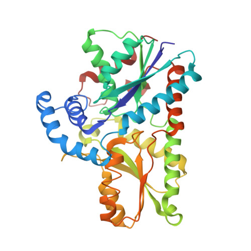
    Ferrochelatase (protoheme ferrolyase, E.C. 4.99.1.1) is the terminal enzyme in heme biosynthesis and catalyzes the insertion of ferrous iron into protoporphyrin IX to form protoheme IX (heme). Within the past two years, X-ray crystallographic data obtained with human ferrochelatase have clearly shown that significant structural changes occur during catalysis that are predicted to facilitate metal insertion and product release. One unanswered question about ferrochelatase involves defining the mechanism whereby some metals, such as divalent Fe, Co, Ni, and Zn, can be used by the enzyme in vitro to produce the corresponding metalloporphyrins, while other metals, such as divalent Mn, Hg, Cd, or Pb, are inhibitors of the enzyme. Through the use of high-resolution X-ray crystallography along with characterization of metal species via their anomalous diffraction, the identity and position of Hg, Cd, Ni, or Mn in the center of enzyme-bound porphyrin macrocycle were determined. When Pb, Hg, Cd, or Ni was present in the macrocycle, the conserved pi helix was in the extended, partially unwound "product release" state. Interestingly, in the structure of ferrochelatase with Mn-porphyrin bound, the pi helix is not extended or unwound and is in the "substrate-bound" conformation. These findings show that at least in the cases of Mn, Pb, Cd, and Hg, metal "inhibition" of ferrochelatase is not due to the inability of the enzyme to insert the metal into the macrocycle or by binding to a second metal binding site as has been previously proposed. Rather, inhibition occurs after metal insertion and results from poor or diminished product release. Possible explanations for the lack of product release are proposed herein.


  • Organizational Affiliation
    • Biomedical and Health Sciences Institute, Department of Biochemistry and Molecular Biology, University of Georgia, Athens, 30602, USA.

Macromolecule Content 

  • Total Structure Weight: 85.55 kDa 
  • Atom Count: 6,583 
  • Modeled Residue Count: 718 
  • Deposited Residue Count: 718 
  • Unique protein chains: 1

Macromolecules

Find similar proteins by:|  3D Structure
Entity ID: 1
MoleculeChains  Sequence LengthOrganismDetailsImage
Ferrochelatase, mitochondrial
A, B
359Homo sapiensMutation(s): 1 
Gene Names: FECH
EC: 4.99.1.1 (PDB Primary Data), 4.98.1.1 (UniProt)
UniProt & NIH Common Fund Data Resources
Find proteins for P22830 (Homo sapiens)
Explore P22830 
Go to UniProtKB:  P22830
PHAROS:  P22830
GTEx:  ENSG00000066926 
Entity Groups
Sequence Clusters30% Identity50% Identity70% Identity90% Identity95% Identity100% Identity
UniProt GroupP22830
Sequence Annotations
Expand
Reference Sequence

Small Molecules

Ligands 5 Unique
IDChains Name / Formula / InChI Key2D Diagram3D Interactions
FDE

Query on FDE


Download:Ideal Coordinates CCD File
H [auth A],
M [auth B]
FE(III) DEUTEROPORPHYRIN IX
C30 H28 Fe N4 O4
QQYZTXBVPVYDJC-RWRCOHKGSA-L
CHD

Query on CHD


Download:Ideal Coordinates CCD File
D [auth A],
E [auth A],
J [auth B],
K [auth B]
CHOLIC ACID
C24 H40 O5
BHQCQFFYRZLCQQ-OELDTZBJSA-N
FES

Query on FES


Download:Ideal Coordinates CCD File
C [auth A],
I [auth B]
FE2/S2 (INORGANIC) CLUSTER
Fe2 S2
NIXDOXVAJZFRNF-UHFFFAOYSA-N
GOL

Query on GOL


Download:Ideal Coordinates CCD File
F [auth A]GLYCEROL
C3 H8 O3
PEDCQBHIVMGVHV-UHFFFAOYSA-N
IMD

Query on IMD


Download:Ideal Coordinates CCD File
G [auth A],
L [auth B]
IMIDAZOLE
C3 H5 N2
RAXXELZNTBOGNW-UHFFFAOYSA-O

Experimental Data & Validation

Experimental Data

  • Method: X-RAY DIFFRACTION
  • Resolution: 2.00 Å
  • R-Value Free:  0.201 (Depositor), 0.176 (DCC) 
  • R-Value Work:  0.170 (Depositor), 0.173 (DCC) 
  • R-Value Observed: 0.172 (Depositor) 
Space Group: P 21 21 21
Unit Cell:
Length ( Å )Angle ( ˚ )
a = 87.574α = 90
b = 93.687β = 90
c = 109.483γ = 90
Software Package:
Software NamePurpose
REFMACrefinement
CNSrefinement
ADSCdata collection
HKL-2000data reduction
SCALEPACKdata scaling
CNSphasing

Structure Validation

View Full Validation Report


Ligand Structure Quality Assessment 


Entry History 

Deposition Data

Revision History  (Full details and data files)

  • Version 1.0: 2009-11-10
    Type: Initial release
  • Version 1.1: 2011-07-13
    Changes: Non-polymer description, Version format compliance
  • Version 1.2: 2019-07-24
    Changes: Data collection, Refinement description
  • Version 1.3: 2024-02-21
    Changes: Data collection, Database references, Derived calculations