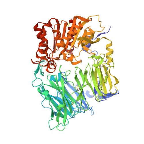
3HAC

The structure of DPP-4 in complex with piperidine fused imidazopyridine 34


Experimental Data Snapshot

  • Method: X-RAY DIFFRACTION
  • Resolution: 2.00 Å
  • R-Value Free: 0.205 
  • R-Value Work: 0.171 
  • R-Value Observed: 0.173 

Starting Model: experimental
View more details

wwPDB Validation   3D Report Full Report


Ligand Structure Quality Assessment 


This is version 3.2 of the entry. See complete history


Literature

Aminopiperidine-fused imidazoles as dipeptidyl peptidase-IV inhibitors

Edmondson, S.D.Mastracchio, A.Cox, J.M.Eiermann, G.J.He, H.Lyons, K.A.Patel, R.A.Patel, S.B.Petrov, A.Scapin, G.Wu, J.K.Xu, S.Zhu, B.Thornberry, N.A.Roy, R.S.Weber, A.E.

(2009) Bioorg Med Chem Lett 19: 4097-4101

  • DOI: https://doi.org/10.1016/j.bmcl.2009.06.011
  • Primary Citation of Related Structures:  
    3HAB, 3HAC

  • PubMed Abstract: 

    A new series of DPP-4 inhibitors derived from piperidine-fused benzimidazoles and imidazopyridines is described. Optimization of this class of DPP-4 inhibitors led to the discovery of imidazopyridine 34. The potency, selectivity, cross-species DMPK profiles, and in vivo efficacy of 34 is reported.


  • Organizational Affiliation

    Department of Medicinal Chemistry, Merck & Co. Inc., PO Box 2000, Rahway, NJ 07065, USA. scott_edmondson@merck.com


Macromolecules
Find similar proteins by:  (by identity cutoff)  |  3D Structure
Entity ID: 1
MoleculeChains Sequence LengthOrganismDetailsImage
Dipeptidyl peptidase 4 soluble form
A, B
728Homo sapiensMutation(s): 1 
Gene Names: DPP4ADCP2CD26
UniProt & NIH Common Fund Data Resources
Find proteins for P27487 (Homo sapiens)
Explore P27487 
Go to UniProtKB:  P27487
PHAROS:  P27487
GTEx:  ENSG00000197635 
Entity Groups  
Sequence Clusters30% Identity50% Identity70% Identity90% Identity95% Identity100% Identity
UniProt GroupP27487
Glycosylation
Glycosylation Sites: 6Go to GlyGen: P27487-1
Sequence Annotations
Expand
  • Reference Sequence
Oligosaccharides

Help

Entity ID: 2
MoleculeChains Length2D Diagram Glycosylation3D Interactions
2-acetamido-2-deoxy-beta-D-glucopyranose-(1-4)-2-acetamido-2-deoxy-beta-D-glucopyranose
C, D, E, F, G
C, D, E, F, G, H, I, J, K
2N-Glycosylation
Glycosylation Resources
GlyTouCan:  G42666HT
GlyCosmos:  G42666HT
GlyGen:  G42666HT
Small Molecules
Binding Affinity Annotations 
IDSourceBinding Affinity
361 BindingDB:  3HAC IC50: min: 8.3, max: 67 (nM) from 3 assay(s)
EC50: 112 (nM) from 1 assay(s)
PDBBind:  3HAC IC50: 6.7 (nM) from 1 assay(s)
Experimental Data & Validation

Experimental Data

  • Method: X-RAY DIFFRACTION
  • Resolution: 2.00 Å
  • R-Value Free: 0.205 
  • R-Value Work: 0.171 
  • R-Value Observed: 0.173 
  • Space Group: P 21 21 21
Unit Cell:
Length ( Å )Angle ( ˚ )
a = 118.12α = 90
b = 125.496β = 90
c = 136.902γ = 90
Software Package:
Software NamePurpose
ADSCdata collection
REFMACrefinement
HKL-2000data reduction
HKL-2000data scaling
REFMACphasing

Structure Validation

View Full Validation Report


Ligand Structure Quality Assessment 


Entry History 

Deposition Data

  • Released Date: 2009-07-21 
  • Deposition Author(s): Scapin, G.

Revision History  (Full details and data files)

  • Version 1.0: 2009-07-21
    Type: Initial release
  • Version 1.1: 2011-07-13
    Changes: Advisory, Version format compliance
  • Version 2.0: 2020-07-29
    Type: Remediation
    Reason: Carbohydrate remediation
    Changes: Advisory, Atomic model, Data collection, Derived calculations, Structure summary
  • Version 2.1: 2021-10-13
    Changes: Database references, Structure summary
  • Version 3.0: 2021-10-27
    Changes: Advisory, Atomic model, Data collection, Derived calculations, Non-polymer description, Source and taxonomy, Structure summary
  • Version 3.1: 2023-06-21
    Changes: Derived calculations
  • Version 3.2: 2023-09-20
    Changes: Data collection, Refinement description