3H9V

Crystal structure of the ATP-gated P2X4 ion channel in the closed, apo state at 3.1 Angstroms


Experimental Data Snapshot

  • Method: X-RAY DIFFRACTION
  • Resolution: 3.10 Å
  • R-Value Free: 0.278 
  • R-Value Work: 0.248 
  • R-Value Observed: 0.249 

wwPDB Validation   3D Report Full Report


This is version 2.1 of the entry. See complete history


Literature

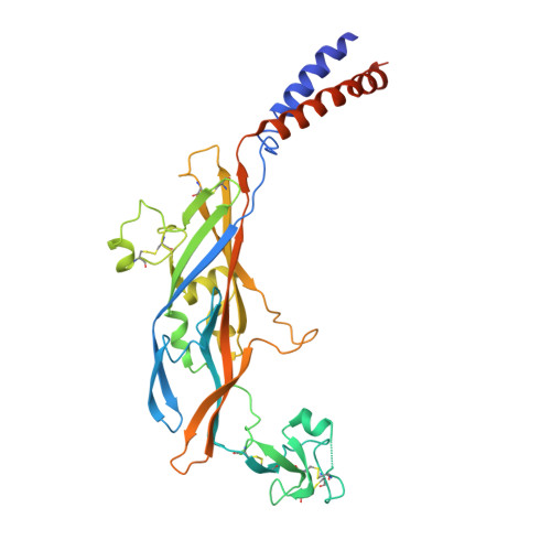
Crystal structure of the ATP-gated P2X(4) ion channel in the closed state.

Kawate, T.Michel, J.C.Birdsong, W.T.Gouaux, E.

(2009) Nature 460: 592-598

  • DOI: https://doi.org/10.1038/nature08198
  • Primary Citation of Related Structures:  
    3H9V, 3I5D

  • PubMed Abstract: 

    P2X receptors are cation-selective ion channels gated by extracellular ATP, and are implicated in diverse physiological processes, from synaptic transmission to inflammation to the sensing of taste and pain. Because P2X receptors are not related to other ion channel proteins of known structure, there is at present no molecular foundation for mechanisms of ligand-gating, allosteric modulation and ion permeation. Here we present crystal structures of the zebrafish P2X(4) receptor in its closed, resting state. The chalice-shaped, trimeric receptor is knit together by subunit-subunit contacts implicated in ion channel gating and receptor assembly. Extracellular domains, rich in beta-strands, have large acidic patches that may attract cations, through fenestrations, to vestibules near the ion channel. In the transmembrane pore, the 'gate' is defined by an approximately 8 A slab of protein. We define the location of three non-canonical, intersubunit ATP-binding sites, and suggest that ATP binding promotes subunit rearrangement and ion channel opening.


  • Organizational Affiliation

    Vollum Institute, Oregon Health and Science University, 3181 Southwest Sam Jackson Park Road, Oregon 97239, USA.


Macromolecules
Find similar proteins by:  (by identity cutoff)  |  3D Structure
Entity ID: 1
MoleculeChains Sequence LengthOrganismDetailsImage
P2X purinoceptor356Danio rerioMutation(s): 4 
Gene Names: p2rx4aP2X4.1
Membrane Entity: Yes 
UniProt
Find proteins for Q6NYR1 (Danio rerio)
Explore Q6NYR1 
Go to UniProtKB:  Q6NYR1
Entity Groups  
Sequence Clusters30% Identity50% Identity70% Identity90% Identity95% Identity100% Identity
UniProt GroupQ6NYR1
Sequence Annotations
Expand
  • Reference Sequence
Oligosaccharides

Help

Entity ID: 2
MoleculeChains Length2D Diagram Glycosylation3D Interactions
2-acetamido-2-deoxy-beta-D-glucopyranose-(1-4)-2-acetamido-2-deoxy-beta-D-glucopyranose
B, C
2N-Glycosylation
Glycosylation Resources
GlyTouCan:  G42666HT
GlyCosmos:  G42666HT
GlyGen:  G42666HT
Experimental Data & Validation

Experimental Data

  • Method: X-RAY DIFFRACTION
  • Resolution: 3.10 Å
  • R-Value Free: 0.278 
  • R-Value Work: 0.248 
  • R-Value Observed: 0.249 
  • Space Group: H 3 2
Unit Cell:
Length ( Å )Angle ( ˚ )
a = 100.836α = 90
b = 100.836β = 90
c = 431.316γ = 120
Software Package:
Software NamePurpose
DENZOdata reduction
SCALEPACKdata scaling
PHENIXrefinement
PDB_EXTRACTdata extraction
ADSCdata collection
HKL-2000data reduction
PHASERphasing

Structure Validation

View Full Validation Report


Entry History 

Deposition Data

Revision History  (Full details and data files)

  • Version 1.0: 2009-07-28
    Type: Initial release
  • Version 1.1: 2011-07-13
    Changes: Non-polymer description, Version format compliance
  • Version 1.2: 2017-11-01
    Changes: Refinement description
  • Version 2.0: 2020-07-29
    Type: Remediation
    Reason: Carbohydrate remediation
    Changes: Advisory, Atomic model, Data collection, Database references, Derived calculations, Structure summary
  • Version 2.1: 2021-10-13
    Changes: Database references, Structure summary