A combined crystallographic and molecular dynamics study of cathepsin L retrobinding inhibitors
Shenoy, R.T., Chowdhury, S.F., Kumar, S., Joseph, L., Purisima, E.O., Sivaraman, J.(2009) J Med Chem 52: 6335-6346
- PubMed: 19761244 Search on PubMed
- DOI: https://doi.org/10.1021/jm900596y
- Primary Citation Related Structures:
3H89, 3H8B, 3H8C - PubMed Abstract:
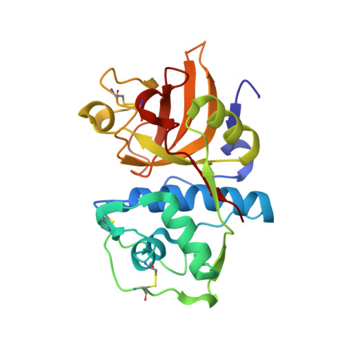
We report the crystal structures of three noncovalent retrobinding inhibitors in complex with mature cathepsin L up to resolutions of 2.5, 1.8, and 2.5 A, respectively. These inhibitors were Bpa-(Nepsilon-Bpa)Lys-DArg-Tyr-Npe, Bpa-(Nepsilon-Bpa)Lys-DArg-Phe-Npe, and Bpa-MCys-DArg-Phe-Npe, where Bpa = biphenylacetyl and Pea = N-phenylethyl. These were selected to clarify the binding mode of the biphenyl groups in the S' subsites because the addition of a second biphenyl does not improve potency. Examination of the symmetry-related monomers in the crystal structures revealed inhibitor-inhibitor crystal packing interactions. Molecular dynamics simulations were then used to explore the structure and dynamical behavior of the isolated protein-ligand complexes in solution. In the simulations, the backbone biphenyl groups for all three inhibitors ended up in the same location despite having started out in different orientations in the initial crystal structure conformations. The lack of improved potency of the larger inhibitors over the smaller one is attributed to a correspondingly greater entropic cost of binding.
- Department of Biological Sciences, National University of Singapore, 14 Science Drive 4, Singapore 117543.
Organizational Affiliation: