3H61

Catalytic domain of human Serine/Threonine Phosphatase 5 (PP5c) with two Mn2+ atoms originally soaked with norcantharidin (which is present in the structure in the hydrolyzed form)


Experimental Data Snapshot

  • Method: X-RAY DIFFRACTION
  • Resolution: 1.45 Å
  • R-Value Free: 0.194 
  • R-Value Work: 0.161 
  • R-Value Observed: 0.164 

wwPDB Validation   3D Report Full Report


Ligand Structure Quality Assessment 


This is version 1.2 of the entry. See complete history


Literature

Structural basis of serine/threonine phosphatase inhibition by the archetypal small molecules cantharidin and norcantharidin

Bertini, I.Calderone, V.Fragai, M.Luchinat, C.Talluri, E.

(2009) J Med Chem 52: 4838-4843

  • DOI: https://doi.org/10.1021/jm900610k
  • Primary Citation of Related Structures:  
    3H60, 3H61, 3H62, 3H63, 3H64, 3H66, 3H67, 3H68, 3H69

  • PubMed Abstract: 

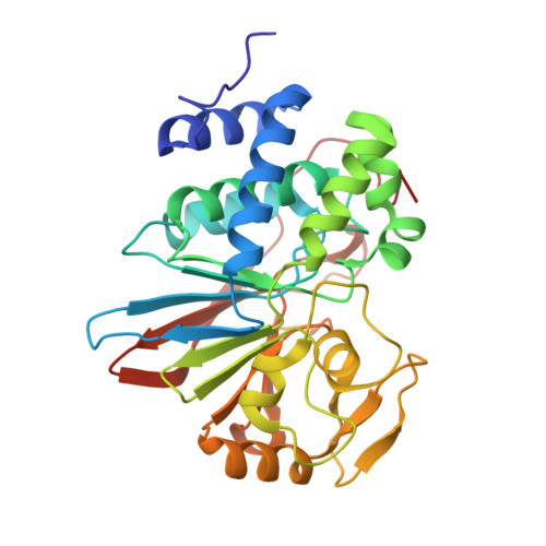
    The inhibition of a subgroup of human serine/threonine protein phosphatases is responsible for the cytotoxicity of cantharidin and norcantharidin against tumor cells. It is shown that the anhydride rings of cantharidin and norcantharidin are hydrolyzed when bound to the catalytic domain of the human serine/threonine protein phosphatases 5 (PP5c), and the high-resolution crystal structures of PP5c complexed with the corresponding dicarboxylic acid derivatives of the two molecules are reported. Norcantharidin shows a unique binding conformation with the catalytically active Mn2PP5c, while cantharidin is characterized by a double conformation in its binding mode to the protein. Different binding modes of norcantharidin are observed depending of whether the starting ligand is in the anhydride or in the dicarboxylic acid form. All these structures will provide the basis for the rational design of new cantharidin-based drugs.


  • Organizational Affiliation

    Department of Chemistry, University of Florence, Via della Lastruccia 3, 50019 Sesto Fiorentino, Italy. bertini@cerm.unifi.it


Macromolecules
Find similar proteins by:  (by identity cutoff)  |  3D Structure
Entity ID: 1
MoleculeChains Sequence LengthOrganismDetailsImage
Serine/threonine-protein phosphatase 5A,
B [auth D]
315Homo sapiensMutation(s): 0 
Gene Names: PPP5CPPP5
EC: 3.1.3.16
UniProt & NIH Common Fund Data Resources
Find proteins for P53041 (Homo sapiens)
Explore P53041 
Go to UniProtKB:  P53041
PHAROS:  P53041
GTEx:  ENSG00000011485 
Entity Groups  
Sequence Clusters30% Identity50% Identity70% Identity90% Identity95% Identity100% Identity
UniProt GroupP53041
Sequence Annotations
Expand
  • Reference Sequence
Experimental Data & Validation

Experimental Data

  • Method: X-RAY DIFFRACTION
  • Resolution: 1.45 Å
  • R-Value Free: 0.194 
  • R-Value Work: 0.161 
  • R-Value Observed: 0.164 
  • Space Group: C 1 2 1
Unit Cell:
Length ( Å )Angle ( ˚ )
a = 154.499α = 90
b = 41.781β = 96.96
c = 105.905γ = 90
Software Package:
Software NamePurpose
ADSCdata collection
MOLREPphasing
REFMACrefinement
MOSFLMdata reduction
SCALAdata scaling

Structure Validation

View Full Validation Report


Ligand Structure Quality Assessment 


Entry History 

Deposition Data

Revision History  (Full details and data files)

  • Version 1.0: 2009-09-29
    Type: Initial release
  • Version 1.1: 2011-07-13
    Changes: Version format compliance
  • Version 1.2: 2023-11-01
    Changes: Data collection, Database references, Derived calculations, Refinement description