3H3C | pdb_00003h3c

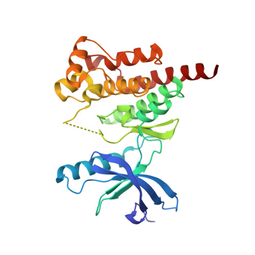
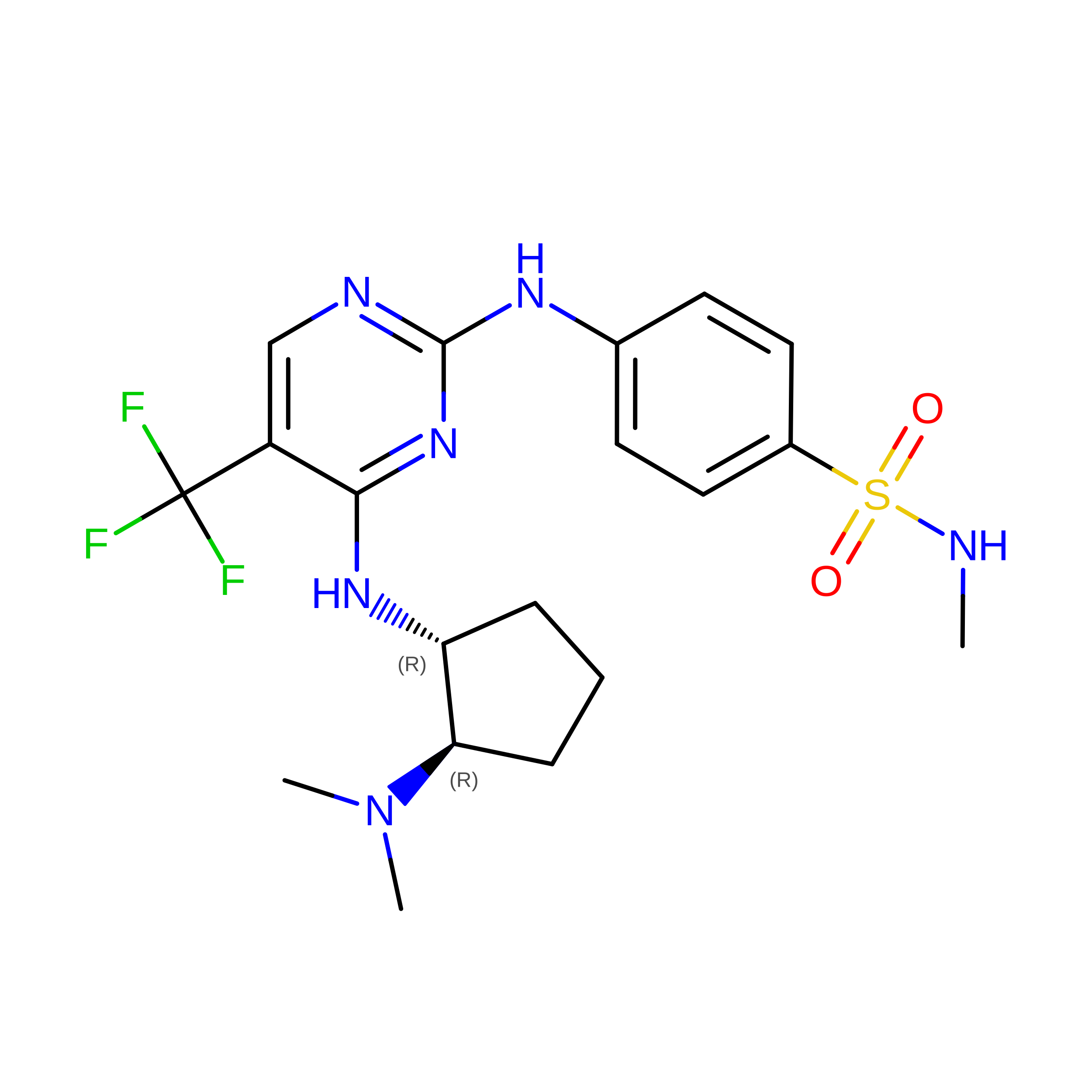
Crystal structure of PYK2 in complex with Sulfoximine-substituted trifluoromethylpyrimidine analog


Experimental Data Snapshot

  • Method: X-RAY DIFFRACTION
  • Resolution: 2.00 Å
  • R-Value Free: 
    0.273 (Depositor), 0.268 (DCC) 
  • R-Value Work: 
    0.222 (Depositor), 0.220 (DCC) 
  • R-Value Observed: 
    0.225 (Depositor) 

wwPDB Validation 3D Report Full Report

Validation slider image for 3H3C

Ligand Structure Quality Assessment 


This is version 1.2 of the entry. See complete history. 

Literature

Sulfoximine-substituted trifluoromethylpyrimidine analogs as inhibitors of proline-rich tyrosine kinase 2 (PYK2) show reduced hERG activity.

Walker, D.P., Zawistoski, M.P., McGlynn, M.A., Li, J.C., Kung, D.W., Bonnette, P.C., Baumann, A., Buckbinder, L., Houser, J.A., Boer, J., Mistry, A., Han, S., Xing, L., Guzman-Perez, A.

(2009) Bioorg Med Chem Lett 19: 3253-3258

  • DOI: https://doi.org/10.1016/j.bmcl.2009.04.093
  • Primary Citation Related Structures: 
    3H3C

  • PubMed Abstract: 

    The synthesis, in vitro properties, and in vivo pharmacokinetics for a series of sulfoximine-substituted trifluoromethylpyrimidines as inhibitors of proline-rich tyrosine kinase, a target for the possible treatment of osteoporosis, are described. These compounds were prepared as surrogates of the corresponding sulfone compound 1. Sulfone 1 was an attractive PYK2 lead compound; however, subsequent studies determined this compound possessed high dofetilide binding, which is an early indicator of cardiovascular safety. Surprisingly, the corresponding sulfoximine analogs displayed significantly lower dofetilide binding, which, for N-methylsulfoximine (S)-14a, translated to lower activity in a patch clamp hERG K(+) ion channel screen. In addition, compound (S)-14a shows good oral exposure in a rat pharmacokinetic model.


  • Organizational Affiliation: 
    • Pfizer Global Research and Development, Eastern Point Road, Groton, CT 06340, USA. daniel.p.walker@pfizer.com

Macromolecule Content 

  • Total Structure Weight: 33.13 kDa 
  • Atom Count: 2,219 
  • Modeled Residue Count: 263 
  • Deposited Residue Count: 277 
  • Unique protein chains: 1

Macromolecules

Find similar proteins by:|  3D Structure
Entity ID: 1
MoleculeChains  Sequence LengthOrganismDetailsImage
Protein tyrosine kinase 2 beta277Homo sapiensMutation(s): 0 
Gene Names: PTK2B, FAK2, PYK2, RAFTK
EC: 2.7.10.2
UniProt & NIH Common Fund Data Resources
Find proteins for Q14289 (Homo sapiens)
Explore Q14289 
Go to UniProtKB:  Q14289
PHAROS:  Q14289
GTEx:  ENSG00000120899 
Entity Groups
Sequence Clusters30% Identity50% Identity70% Identity90% Identity95% Identity100% Identity
UniProt GroupQ14289
Sequence Annotations
Expand
Reference Sequence

Experimental Data & Validation

Experimental Data

  • Method: X-RAY DIFFRACTION
  • Resolution: 2.00 Å
  • R-Value Free:  0.273 (Depositor), 0.268 (DCC) 
  • R-Value Work:  0.222 (Depositor), 0.220 (DCC) 
  • R-Value Observed: 0.225 (Depositor) 
Space Group: P 1 21 1
Unit Cell:
Length ( Å )Angle ( ˚ )
a = 37.281α = 90
b = 92.83β = 92.64
c = 42.843γ = 90
Software Package:
Software NamePurpose
HKL-2000data collection
REFMACrefinement
HKL-2000data reduction
HKL-2000data scaling
REFMACphasing

Structure Validation

View Full Validation Report


Ligand Structure Quality Assessment 


Entry History 

Deposition Data

Revision History  (Full details and data files)

  • Version 1.0: 2009-05-26
    Type: Initial release
  • Version 1.1: 2011-07-13
    Changes: Version format compliance
  • Version 1.2: 2024-02-21
    Changes: Data collection, Database references, Derived calculations