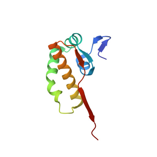
Structure of the C-terminal domain of nsp4 from feline coronavirus
Manolaridis, I., Wojdyla, J.A., Panjikar, S., Snijder, E.J., Gorbalenya, A.E., Berglind, H., Nordlund, P., Coutard, B., Tucker, P.A.(2009) Acta Crystallogr D Biol Crystallogr 65: 839-846
- PubMed: 19622868 Search on PubMedSearch on PubMed Central
- DOI: https://doi.org/10.1107/S0907444909018253
- Primary Citation Related Structures:
3GZF - PubMed Abstract:
Coronaviruses are a family of positive-stranded RNA viruses that includes important pathogens of humans and other animals. The large coronavirus genome (26-31 kb) encodes 15-16 nonstructural proteins (nsps) that are derived from two replicase polyproteins by autoproteolytic processing. The nsps assemble into the viral replication-transcription complex and nsp3, nsp4 and nsp6 are believed to anchor this enzyme complex to modified intracellular membranes. The largest part of the coronavirus nsp4 subunit is hydrophobic and is predicted to be embedded in the membranes. In this report, a conserved C-terminal domain ( approximately 100 amino-acid residues) has been delineated that is predicted to face the cytoplasm and has been isolated as a soluble domain using library-based construct screening. A prototypical crystal structure at 2.8 A resolution was obtained using nsp4 from feline coronavirus. Unmodified and SeMet-substituted proteins were crystallized under similar conditions, resulting in tetragonal crystals that belonged to space group P4(3). The phase problem was initially solved by single isomorphous replacement with anomalous scattering (SIRAS), followed by molecular replacement using a SIRAS-derived composite model. The structure consists of a single domain with a predominantly alpha-helical content displaying a unique fold that could be engaged in protein-protein interactions.
- EMBL Hamburg Outstation, D-22603 Hamburg, Germany.
Organizational Affiliation: