3GYW | pdb_00003gyw

Crystal structure of nucleosome assembly protein from Plasmodium falciparum at 2.4 A resolution


Experimental Data Snapshot

  • Method: X-RAY DIFFRACTION
  • Resolution: 2.40 Å
  • R-Value Free: 
    0.267 (Depositor), 0.264 (DCC) 
  • R-Value Work: 
    0.220 (Depositor), 0.218 (DCC) 

wwPDB Validation 3D Report Full Report

Validation slider image for 3GYW

This is version 1.2 of the entry. See complete history

Literature

Iodide-SAD, SIR and SIRAS phasing for structure solution of a nucleosome assembly protein.

Yogavel, M.Gill, J.Sharma, A.

(2009) Acta Crystallogr D Biol Crystallogr 65: 618-622

  • DOI: https://doi.org/10.1107/S0907444909013171
  • Primary Citation Related Structures: 
    3GYV, 3GYW

  • PubMed Abstract: 

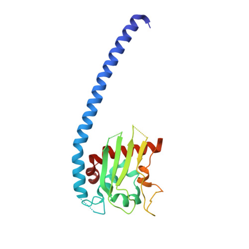
    The crystal structure of Plasmodium falciparum nucleosome assembly protein (PfNapL) was determined by iodide-SAD/SIRAS phasing methods using iodide-SAD data to 3.0 A resolution and native data to 2.4 A resolution. Halide-derivatized PfNapL crystals were obtained using the quick cryo-soaking method in which the native crystals were soaked in a cryosolution consisting of 500 mM NaI for a short period of 30-60 s and data were collected at an in-house X-ray source using Cu Kalpha radiation. Despite a low anomalous signal-to-noise ratio of <1.2 in the >3.5 A resolution bin, the data were sufficient to determine the structure by SAD/SIR/SIRAS methods using the soaked iodides. Previously, structure solution had failed with both molecular-replacement and selenomethionine-derivatization techniques owing to reasons that are detailed in this work. The phasing at low resolution with three iodides per monomer with high temperature factors was successful using any of the SAD, SIR or SIRAS methods.


  • Organizational Affiliation
    • Structural and Computational Biology Group, International Centre for Genetic Engineering and Biotechnology (ICGEB), New Delhi 110 067, India.

Macromolecule Content 

  • Total Structure Weight: 29.67 kDa 
  • Atom Count: 1,584 
  • Modeled Residue Count: 180 
  • Deposited Residue Count: 249 
  • Unique protein chains: 1

Macromolecules

Find similar proteins by:|  3D Structure
Entity ID: 1
MoleculeChains  Sequence LengthOrganismDetailsImage
Nucleosome assembly protein 1, putative249Plasmodium falciparum 3D7Mutation(s): 0 
Gene Names: pfl0185c
UniProt
Find proteins for Q8I608 (Plasmodium falciparum (isolate 3D7))
Explore Q8I608 
Go to UniProtKB:  Q8I608
Entity Groups
Sequence Clusters30% Identity50% Identity70% Identity90% Identity95% Identity100% Identity
UniProt GroupQ8I608
Sequence Annotations
Expand
Reference Sequence

Experimental Data & Validation

Experimental Data

  • Method: X-RAY DIFFRACTION
  • Resolution: 2.40 Å
  • R-Value Free:  0.267 (Depositor), 0.264 (DCC) 
  • R-Value Work:  0.220 (Depositor), 0.218 (DCC) 
Space Group: C 1 2 1
Unit Cell:
Length ( Å )Angle ( ˚ )
a = 76.24α = 90
b = 37.87β = 99.7
c = 79.14γ = 90
Software Package:
Software NamePurpose
MAR345dtbdata collection
PHENIXmodel building
REFMACrefinement
HKL-2000data reduction
HKL-2000data scaling
PHENIXphasing

Structure Validation

View Full Validation Report


Entry History 

Deposition Data

Revision History  (Full details and data files)

  • Version 1.0: 2009-05-12
    Type: Initial release
  • Version 1.1: 2011-07-13
    Changes: Source and taxonomy, Version format compliance
  • Version 1.2: 2024-03-20
    Changes: Data collection, Database references