3GWZ

Structure of the Mitomycin 7-O-methyltransferase MmcR


Experimental Data Snapshot

  • Method: X-RAY DIFFRACTION
  • Resolution: 1.91 Å
  • R-Value Free: 0.210 
  • R-Value Work: 0.174 
  • R-Value Observed: 0.176 

wwPDB Validation   3D Report Full Report


Ligand Structure Quality Assessment 


This is version 1.2 of the entry. See complete history


Literature

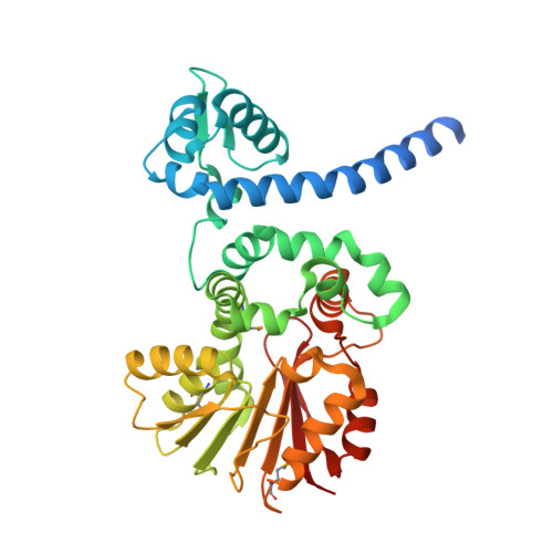
Structural characterization of the mitomycin 7-O-methyltransferase.

Singh, S.Chang, A.Goff, R.D.Bingman, C.A.Gruschow, S.Sherman, D.H.Phillips, G.N.Thorson, J.S.

(2011) Proteins 79: 2181-2188

  • DOI: https://doi.org/10.1002/prot.23040
  • Primary Citation of Related Structures:  
    3GWZ, 3GXO

  • PubMed Abstract: 
  • Mitomycins are quinone-containing antibiotics, widely used as antitumor drugs in chemotherapy. Mitomycin-7-O-methyltransferase (MmcR), a key tailoring enzyme involved in the biosynthesis of mitomycin in Streptomyces lavendulae, catalyzes the 7-O-methylation of both C9β- and C9α-configured 7-hydroxymitomycins ...

    Mitomycins are quinone-containing antibiotics, widely used as antitumor drugs in chemotherapy. Mitomycin-7-O-methyltransferase (MmcR), a key tailoring enzyme involved in the biosynthesis of mitomycin in Streptomyces lavendulae, catalyzes the 7-O-methylation of both C9β- and C9α-configured 7-hydroxymitomycins. We have determined the crystal structures of the MmcR-S-adenosylhomocysteine (SAH) binary complex and MmcR-SAH-mitomycin A (MMA) ternary complex at resolutions of 1.9and 2.3 Å, respectively. The study revealed MmcR to adopt a common S-adenosyl-L-methionine-dependent O-methyltransferase fold and the presence of a structurally conserved active site general acid-base pair is consistent with a proton-assisted methyltransfer common to most methyltransferases. Given the importance of C7 alkylation to modulate mitomycin redox potential, this study may also present a template toward the future engineering of catalysts to generate uniquely bioactive mitomycins.


    Organizational Affiliation

    Division of Pharmaceutical Sciences, Wisconsin Center for Natural Product Research, School of Pharmacy, University of Wisconsin-Madison, Madison, Wisconsin 53705, USA.


Macromolecules
Find similar proteins by:  (by identity cutoff)  |  3D Structure
Entity ID: 1
MoleculeChainsSequence LengthOrganismDetailsImage
MmcRA,
B [auth D],
C,
D [auth B]
369Streptomyces lavendulaeMutation(s): 0 
Gene Names: mmcR
EC: 2.1.1.316
UniProt
Find proteins for Q9X5T6 (Streptomyces lavendulae)
Explore Q9X5T6 
Go to UniProtKB:  Q9X5T6
Entity Groups  
Sequence Clusters30% Identity50% Identity70% Identity90% Identity95% Identity100% Identity
UniProt GroupQ9X5T6
Protein Feature View
Expand
  • Reference Sequence
Small Molecules
Modified Residues  1 Unique
IDChainsTypeFormula2D DiagramParent
MSE
Query on MSE
A,
B [auth D],
C,
D [auth B]
L-PEPTIDE LINKINGC5 H11 N O2 SeMET
Experimental Data & Validation

Experimental Data

  • Method: X-RAY DIFFRACTION
  • Resolution: 1.91 Å
  • R-Value Free: 0.210 
  • R-Value Work: 0.174 
  • R-Value Observed: 0.176 
  • Space Group: P 21 21 21
Unit Cell:
Length ( Å )Angle ( ˚ )
a = 88.372α = 90
b = 98.918β = 90
c = 171.119γ = 90
Software Package:
Software NamePurpose
DENZOdata reduction
SCALEPACKdata scaling
SHARPphasing
DMphasing
PHENIXrefinement
PDB_EXTRACTdata extraction
HKL-2000data reduction
HKL-2000data scaling

Structure Validation

View Full Validation Report


Ligand Structure Quality Assessment 


Entry History 

Deposition Data

Revision History  (Full details and data files)

  • Version 1.0: 2010-04-07
    Type: Initial release
  • Version 1.1: 2011-07-13
    Changes: Version format compliance
  • Version 1.2: 2017-11-01
    Changes: Advisory, Refinement description