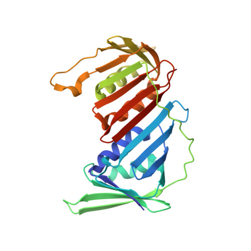
A charged residue at the subunit interface of PCNA promotes trimer formation by destabilizing alternate subunit interactions.
Freudenthal, B.D., Gakhar, L., Ramaswamy, S., Washington, M.T.(2009) Acta Crystallogr D Biol Crystallogr 65: 560-566
- PubMed: 19465770
- DOI: https://doi.org/10.1107/S0907444909011329
- Primary Citation of Related Structures:
3GPM, 3GPN - PubMed Abstract:
Eukaryotic proliferating cell nuclear antigen (PCNA) is an essential replication accessory factor that interacts with a variety of proteins involved in DNA replication and repair. Each monomer of PCNA has an N-terminal domain A and a C-terminal domain B. In the structure of the wild-type PCNA protein, domain A of one monomer interacts with domain B of a neighboring monomer to form a ring-shaped trimer. Glu113 is a conserved residue at the subunit interface in domain A. Two distinct X-ray crystal structures have been determined of a mutant form of PCNA with a substitution at this position (E113G) that has previously been studied because of its effect on translesion synthesis. The first structure was the expected ring-shaped trimer. The second structure was an unanticipated nontrimeric form of the protein. In this nontrimeric form, domain A of one PCNA monomer interacts with domain A of a neighboring monomer, while domain B of this monomer interacts with domain B of a different neighboring monomer. The B-B interface is stabilized by an antiparallel beta-sheet and appears to be structurally similar to the A-B interface observed in the trimeric form of PCNA. The A-A interface, in contrast, is primarily stabilized by hydrophobic interactions. Because the E113G substitution is located on this hydrophobic surface, the A-A interface should be less favorable in the case of the wild-type protein. This suggests that the side chain of Glu113 promotes trimer formation by destabilizing these possible alternate subunit interactions.
- Department of Biochemistry, University of Iowa College of Medicine, Iowa City, IA 52242-1109, USA.
Organizational Affiliation:


