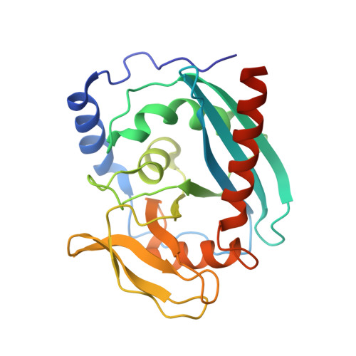
Crystal structure of the Endonuclease V (SAV1684) from Streptomyces avermitilis. Northeast Structural Genomics Consortium Target SvR196.
Forouhar, F., Abashidze, M., Hussain, M., Seetharaman, J., Fang, F., Xiao, R., Cunningham, K., Ma, L., Owens, L., Chen, C.X., Everett, J.K., Nair, R., Acton, T.B., Rost, B., Montelione, G.T., Tong, L., Hunt, J.F., Northeast Structural Genomics Consortium (NESG)To be published.