3GH8

Crystal structure of Mus musculus iodotyrosine deiodinase (IYD) bound to FMN and di-iodotyrosine (DIT)


Experimental Data Snapshot

  • Method: X-RAY DIFFRACTION
  • Resolution: 2.61 Å
  • R-Value Free: 0.264 
  • R-Value Work: 0.181 
  • R-Value Observed: 0.185 

wwPDB Validation   3D Report Full Report


Ligand Structure Quality Assessment 


This is version 1.2 of the entry. See complete history


Literature

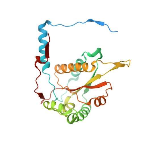
Crystal structure of iodotyrosine deiodinase, a novel flavoprotein responsible for iodide salvage in thyroid glands.

Thomas, S.R.McTamney, P.M.Adler, J.M.Laronde-Leblanc, N.Rokita, S.E.

(2009) J Biol Chem 284: 19659-19667

  • DOI: https://doi.org/10.1074/jbc.M109.013458
  • Primary Citation of Related Structures:  
    3GB5, 3GFD, 3GH8

  • PubMed Abstract: 

    The flavoprotein iodotyrosine deiodinase (IYD) salvages iodide from mono- and diiodotyrosine formed during the biosynthesis of the thyroid hormone thyroxine. Expression of a soluble domain of this membrane-bound enzyme provided sufficient material for crystallization and characterization by x-ray diffraction. The structures of IYD and two co-crystals containing substrates, mono- and diiodotyrosine, alternatively, were solved at resolutions of 2.0, 2.45, and 2.6 A, respectively. The structure of IYD is homologous to others in the NADH oxidase/flavin reductase superfamily, but the position of the active site lid in IYD defines a new subfamily within this group that includes BluB, an enzyme associated with vitamin B(12) biosynthesis. IYD and BluB also share key interactions involving their bound flavin mononucleotide that suggest a unique catalytic behavior within the superfamily. Substrate coordination to IYD induces formation of an additional helix and coil that act as an active site lid to shield the resulting substrate.flavin complex from solvent. This complex is stabilized by aromatic stacking and extensive hydrogen bonding between the substrate and flavin. The carbon-iodine bond of the substrate is positioned directly over the C-4a/N-5 region of the flavin to promote electron transfer. These structures now also provide a molecular basis for understanding thyroid disease based on mutations of IYD.


  • Organizational Affiliation

    Department of Chemistry and Biochemistry, Center for Biomolecular Structure and Organization, University of Maryland, College Park, Maryland 20742, USA.


Macromolecules
Find similar proteins by:  (by identity cutoff)  |  3D Structure
Entity ID: 1
MoleculeChains Sequence LengthOrganismDetailsImage
Iodotyrosine dehalogenase 1
A, B, C, D, E
A, B, C, D, E, F, G, H
259Mus musculusMutation(s): 0 
Gene Names: Dehal1Iyd
UniProt
Find proteins for Q9DCX8 (Mus musculus)
Explore Q9DCX8 
Go to UniProtKB:  Q9DCX8
Entity Groups  
Sequence Clusters30% Identity50% Identity70% Identity90% Identity95% Identity100% Identity
UniProt GroupQ9DCX8
Sequence Annotations
Expand
  • Reference Sequence
Small Molecules
Ligands 3 Unique
IDChains Name / Formula / InChI Key2D Diagram3D Interactions
FMN
Query on FMN

Download Ideal Coordinates CCD File 
I [auth A]
K [auth B]
M [auth C]
P [auth D]
R [auth E]
I [auth A],
K [auth B],
M [auth C],
P [auth D],
R [auth E],
T [auth F],
V [auth G],
Y [auth H]
FLAVIN MONONUCLEOTIDE
C17 H21 N4 O9 P
FVTCRASFADXXNN-SCRDCRAPSA-N
TYI
Query on TYI

Download Ideal Coordinates CCD File 
J [auth A]
L [auth B]
N [auth C]
Q [auth D]
S [auth E]
J [auth A],
L [auth B],
N [auth C],
Q [auth D],
S [auth E],
U [auth F],
W [auth G],
Z [auth H]
3,5-DIIODOTYROSINE
C9 H9 I2 N O3
NYPYHUZRZVSYKL-ZETCQYMHSA-N
PO4
Query on PO4

Download Ideal Coordinates CCD File 
O [auth C],
X [auth G]
PHOSPHATE ION
O4 P
NBIIXXVUZAFLBC-UHFFFAOYSA-K
Experimental Data & Validation

Experimental Data

  • Method: X-RAY DIFFRACTION
  • Resolution: 2.61 Å
  • R-Value Free: 0.264 
  • R-Value Work: 0.181 
  • R-Value Observed: 0.185 
  • Space Group: P 1 21 1
Unit Cell:
Length ( Å )Angle ( ˚ )
a = 50.61α = 90
b = 112.567β = 89.92
c = 189.253γ = 90
Software Package:
Software NamePurpose
REFMACrefinement
PDB_EXTRACTdata extraction
HKL-2000data collection
HKL-2000data reduction
HKL-2000data scaling
MOLREPphasing

Structure Validation

View Full Validation Report


Ligand Structure Quality Assessment 


Entry History 

Deposition Data

Revision History  (Full details and data files)

  • Version 1.0: 2009-05-12
    Type: Initial release
  • Version 1.1: 2011-07-13
    Changes: Advisory, Version format compliance
  • Version 1.2: 2023-09-06
    Changes: Data collection, Database references, Derived calculations, Refinement description