3GGS | pdb_00003ggs

Human purine nucleoside phosphorylase double mutant E201Q,N243D complexed with 2-fluoro-2'-deoxyadenosine


Experimental Data Snapshot

  • Method: X-RAY DIFFRACTION
  • Resolution: 2.52 Å
  • R-Value Free: 
    0.251 (Depositor) 
  • R-Value Work: 
    0.212 (Depositor) 
  • R-Value Observed: 
    0.214 (Depositor) 

Starting Model: experimental
View more details

wwPDB Validation 3D Report Full Report

Validation slider image for 3GGS

Ligand Structure Quality Assessment 


This is version 1.4 of the entry. See complete history

Literature

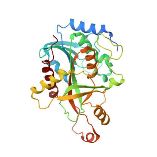
Structure of a mutant human purine nucleoside phosphorylase with the prodrug, 2-fluoro-2'-deoxyadenosine and the cytotoxic drug, 2-fluoroadenine.

Afshar, S.Sawaya, M.R.Morrison, S.L.

(2009) Protein Sci 18: 1107-1114

  • DOI: https://doi.org/10.1002/pro.91
  • Primary Citation Related Structures: 
    3GB9, 3GGS

  • PubMed Abstract: 

    A double mutant of human purine nucleoside phosphorylase (hDM) with the amino acid mutations Glu201Gln:Asn243Asp cleaves adenosine-based prodrugs to their corresponding cytotoxic drugs. When fused to an anti-tumor targeting component, hDM is targeted to tumor cells, where it effectively catalyzes phosphorolysis of the prodrug, 2-fluoro-2'-deoxyadenosine (F-dAdo) to the cytotoxic drug, 2-fluoroadenine (F-Ade). This cytotoxicity should be restricted only to the tumor microenvironment, because the endogenously expressed wild type enzyme cannot use adenosine-based prodrugs as substrates. To gain insight into the interaction of hDM with F-dAdo, we have determined the crystal structures of hDM with F-dAdo and F-Ade. The structures reveal that despite the two mutations, the overall fold of hDM is nearly identical to the wild type enzyme. Importantly, the residues Gln201 and Asp243 introduced by the mutation form hydrogen bond contacts with F-dAdo that result in its binding and catalysis. Comparison of substrate and product complexes suggest that the side chains of Gln201 and Asp243 as well as the purine base rotate during catalysis possibly facilitating cleavage of the glycosidic bond. The two structures suggest why hDM, unlike the wild-type enzyme, can utilize F-dAdo as substrate. More importantly, they provide a critical foundation for further optimization of cleavage of adenosine-based prodrugs, such as F-dAdo by mutants of human purine nucleoside phosphorylase.


  • Organizational Affiliation
    • Department of Microbiology, Immunology, and Molecular Genetics, UCLA-DOE Institute for Genomics and Proteomics, UCLA, Los Angeles, California 90095.

Macromolecule Content 

  • Total Structure Weight: 107.16 kDa 
  • Atom Count: 6,904 
  • Modeled Residue Count: 848 
  • Deposited Residue Count: 933 
  • Unique protein chains: 1

Macromolecules

Find similar proteins by:|  3D Structure
Entity ID: 1
MoleculeChains  Sequence LengthOrganismDetailsImage
Purine nucleoside phosphorylase
A, B, C
311Homo sapiensMutation(s): 2 
Gene Names: NPPNP
EC: 2.4.2.1
UniProt & NIH Common Fund Data Resources
Find proteins for P00491 (Homo sapiens)
Explore P00491 
Go to UniProtKB:  P00491
PHAROS:  P00491
GTEx:  ENSG00000198805 
Entity Groups
Sequence Clusters30% Identity50% Identity70% Identity90% Identity95% Identity100% Identity
UniProt GroupP00491
Sequence Annotations
Expand
Reference Sequence

Small Molecules

Ligands 2 Unique
IDChains Name / Formula / InChI Key2D Diagram3D Interactions
2FD

Query on 2FD


Download:Ideal Coordinates CCD File
D [auth A],
KA [auth C],
O [auth B],
Z [auth C]
5-(6-AMINO-2-FLUORO-PURIN-9-YL)-2-HYDROXYMETHYL-TETRAHYDRO-FURAN-3-OL
C10 H12 F N5 O3
ZWPYUXAXLRFWQC-KVQBGUIXSA-N
SO4

Query on SO4


Download:Ideal Coordinates CCD File
AA [auth C]
BA [auth C]
CA [auth C]
DA [auth C]
E [auth A]
AA [auth C],
BA [auth C],
CA [auth C],
DA [auth C],
E [auth A],
EA [auth C],
F [auth A],
FA [auth C],
G [auth A],
GA [auth C],
H [auth A],
HA [auth C],
I [auth A],
IA [auth C],
J [auth A],
JA [auth C],
K [auth A],
L [auth A],
M [auth A],
N [auth A],
P [auth B],
Q [auth B],
R [auth B],
S [auth B],
T [auth B],
U [auth B],
V [auth B],
W [auth B],
X [auth B],
Y [auth B]
SULFATE ION
O4 S
QAOWNCQODCNURD-UHFFFAOYSA-L

Experimental Data & Validation

Experimental Data

  • Method: X-RAY DIFFRACTION
  • Resolution: 2.52 Å
  • R-Value Free:  0.251 (Depositor) 
  • R-Value Work:  0.212 (Depositor) 
  • R-Value Observed: 0.214 (Depositor) 
Space Group: P 21 21 21
Unit Cell:
Length ( Å )Angle ( ˚ )
a = 71.24α = 90
b = 130.646β = 90
c = 149.433γ = 90
Software Package:
Software NamePurpose
DENZOdata reduction
SCALEPACKdata scaling
REFMACrefinement
PDB_EXTRACTdata extraction
REFMACphasing

Structure Validation

View Full Validation Report


Ligand Structure Quality Assessment 


Entry History 

Deposition Data

Revision History  (Full details and data files)

  • Version 1.0: 2009-04-14
    Type: Initial release
  • Version 1.1: 2011-07-13
    Changes: Advisory, Refinement description, Version format compliance
  • Version 1.2: 2017-11-01
    Changes: Refinement description
  • Version 1.3: 2021-10-20
    Changes: Database references, Derived calculations
  • Version 1.4: 2023-09-06
    Changes: Data collection, Refinement description