3GBR

Anthranilate phosphoribosyl-transferase (TRPD) double mutant D83G F149S from S. solfataricus


Experimental Data Snapshot

  • Method: X-RAY DIFFRACTION
  • Resolution: 2.25 Å
  • R-Value Free: 0.260 
  • R-Value Work: 0.192 
  • R-Value Observed: 0.194 

wwPDB Validation   3D Report Full Report


Ligand Structure Quality Assessment 


This is version 1.5 of the entry. See complete history


Literature

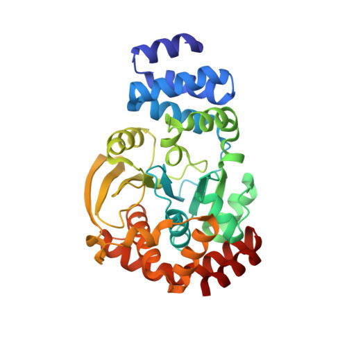
Activation of anthranilate phosphoribosyltransferase from Sulfolobus solfataricus by removal of magnesium inhibition and acceleration of product release

Schlee, S.Deuss, M.Bruning, M.Ivens, A.Schwab, T.Hellmann, N.Mayans, O.Sterner, R.

(2009) Biochemistry 48: 5199-5209

  • DOI: https://doi.org/10.1021/bi802335s
  • Primary Citation of Related Structures:  
    3GBR

  • PubMed Abstract: 

    Anthranilate phosphoribosyltransferase from the hyperthermophilic archaeon Sulfolobus solfataricus (ssAnPRT) is encoded by the sstrpD gene and catalyzes the reaction of anthranilate (AA) with a complex of Mg(2+) and 5'-phosphoribosyl-alpha1-pyrophosphate (Mg.PRPP) to N-(5'-phosphoribosyl)-anthranilate (PRA) and pyrophosphate (PP(i)) within tryptophan biosynthesis. The ssAnPRT enzyme is highly thermostable (half-life at 85 degrees C = 35 min) but only marginally active at ambient temperatures (turnover number at 37 degrees C = 0.33 s(-1)). To understand the reason for the poor catalytic proficiency of ssAnPRT, we have isolated from an sstrpD library the activated ssAnPRT-D83G + F149S double mutant by metabolic complementation of an auxotrophic Escherichia coli strain. Whereas the activity of purified wild-type ssAnPRT is strongly reduced in the presence of high concentrations of Mg(2+) ions, this inhibition is no longer observed in the double mutant and the ssAnPRT-D83G single mutant. The comparison of the crystal structures of activated and wild-type ssAnPRT shows that the D83G mutation alters the binding mode of the substrate Mg.PRPP. Analysis of PRPP and Mg(2+)-dependent enzymatic activity indicates that this leads to a decreased affinity for a second Mg(2+) ion and thus reduces the concentration of enzymes with the inhibitory Mg(2).PRPP complex bound to the active site. Moreover, the turnover number of the double mutant ssAnPRT-D83G + F149S is elevated 40-fold compared to the wild-type enzyme, which can be attributed to an accelerated release of the product PRA. This effect appears to be mainly caused by an increased conformational flexibility induced by the F149S mutation, a hypothesis which is supported by the reduced thermal stability of the ssAnPRT-F149S single mutant.


  • Organizational Affiliation

    Institute of Biophysics and Physical Biochemistry, University of Regensburg, Germany.


Macromolecules
Find similar proteins by:  (by identity cutoff)  |  3D Structure
Entity ID: 1
MoleculeChains Sequence LengthOrganismDetailsImage
Anthranilate phosphoribosyltransferase
A, B
345Saccharolobus solfataricusMutation(s): 2 
Gene Names: trpD
EC: 2.4.2.18
UniProt
Find proteins for P50384 (Saccharolobus solfataricus (strain ATCC 35092 / DSM 1617 / JCM 11322 / P2))
Explore P50384 
Go to UniProtKB:  P50384
Entity Groups  
Sequence Clusters30% Identity50% Identity70% Identity90% Identity95% Identity100% Identity
UniProt GroupP50384
Sequence Annotations
Expand
  • Reference Sequence
Small Molecules
Ligands 4 Unique
IDChains Name / Formula / InChI Key2D Diagram3D Interactions
PRP
Query on PRP

Download Ideal Coordinates CCD File 
C [auth A],
F [auth B]
1-O-pyrophosphono-5-O-phosphono-alpha-D-ribofuranose
C5 H13 O14 P3
PQGCEDQWHSBAJP-TXICZTDVSA-N
PEG
Query on PEG

Download Ideal Coordinates CCD File 
I [auth B],
J [auth B]
DI(HYDROXYETHYL)ETHER
C4 H10 O3
MTHSVFCYNBDYFN-UHFFFAOYSA-N
GOL
Query on GOL

Download Ideal Coordinates CCD File 
K [auth B],
L [auth B]
GLYCEROL
C3 H8 O3
PEDCQBHIVMGVHV-UHFFFAOYSA-N
MN
Query on MN

Download Ideal Coordinates CCD File 
D [auth A],
E [auth A],
G [auth B],
H [auth B]
MANGANESE (II) ION
Mn
WAEMQWOKJMHJLA-UHFFFAOYSA-N
Experimental Data & Validation

Experimental Data

  • Method: X-RAY DIFFRACTION
  • Resolution: 2.25 Å
  • R-Value Free: 0.260 
  • R-Value Work: 0.192 
  • R-Value Observed: 0.194 
  • Space Group: P 1 21 1
Unit Cell:
Length ( Å )Angle ( ˚ )
a = 61.945α = 90
b = 65.993β = 98.81
c = 84.262γ = 90
Software Package:
Software NamePurpose
PHASERphasing
REFMACrefinement
XDSdata reduction
XSCALEdata scaling
XDSdata scaling

Structure Validation

View Full Validation Report


Ligand Structure Quality Assessment 


Entry History 

Deposition Data

Revision History  (Full details and data files)

  • Version 1.0: 2009-12-15
    Type: Initial release
  • Version 1.1: 2011-07-13
    Changes: Non-polymer description, Version format compliance
  • Version 1.2: 2017-11-01
    Changes: Advisory, Refinement description
  • Version 1.3: 2020-07-29
    Type: Remediation
    Reason: Carbohydrate remediation
    Changes: Data collection, Derived calculations, Structure summary
  • Version 1.4: 2021-11-10
    Changes: Advisory, Database references, Structure summary
  • Version 1.5: 2023-11-01
    Changes: Data collection, Refinement description