3G5E | pdb_00003g5e

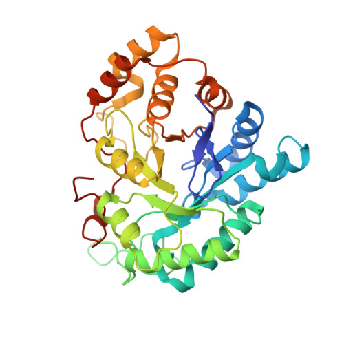
Human aldose reductase complexed with IDD 740 inhibitor


Experimental Data Snapshot

  • Method: X-RAY DIFFRACTION
  • Resolution: 1.80 Å
  • R-Value Free: 
    0.183 (Depositor), 0.182 (DCC) 
  • R-Value Work: 
    0.144 (Depositor), 0.144 (DCC) 
  • R-Value Observed: 
    0.146 (Depositor) 

wwPDB Validation 3D Report Full Report

Validation slider image for 3G5E

Ligand Structure Quality Assessment 


This is version 1.2 of the entry. See complete history. 

Literature

Discovery of [3-(4,5,7-trifluoro-benzothiazol-2-ylmethyl)-pyrrolo[2,3-b]pyridin-1-yl]acetic acids as highly potent and selective inhibitors of aldose reductase for treatment of chronic diabetic complications.

Van Zandt, M.C., Doan, B., Sawicki, D.R., Sredy, J., Podjarny, A.D.

(2009) Bioorg Med Chem Lett 19: 2006-2008

  • DOI: https://doi.org/10.1016/j.bmcl.2009.02.037
  • Primary Citation Related Structures: 
    3G5E

  • PubMed Abstract: 

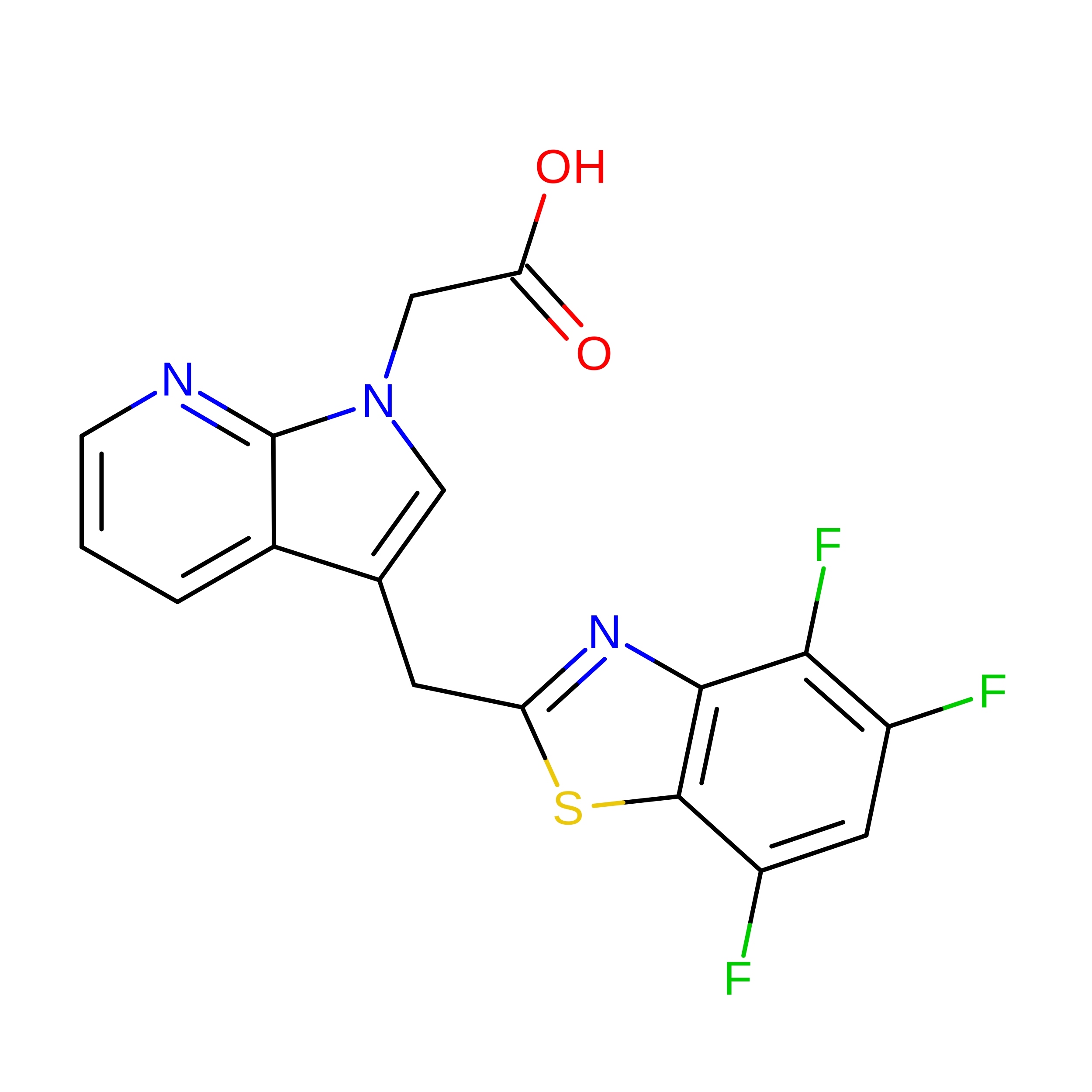
    Efforts to identify treatments for chronic diabetic complications have resulted in the discovery of a novel series of highly potent and selective [3-(4,5,7-trifluoro-benzothiazol-2-ylmethyl)-pyrrolo[2,3-b]pyridin-1-yl]acetic acid aldose reductase inhibitors. The lead candidate, [6-methyl-3-(4,5,7-trifluoro-benzothiazol-2-ylmethyl)-pyrrolo[2,3-b]pyridin-1-yl]acetic acid example 16, inhibits aldose reductase with an IC50 of 8 nM, while being inactive against aldehyde reductase (IC50>100 microM), a related enzyme involved in the detoxification of reactive aldehydes.


  • Organizational Affiliation: 
    • The Institute for Diabetes Discovery, LLC, 23 Business Park Drive, Branford, CT 06405, USA. michael.vanzandt@ipd-discovery.com

Macromolecule Content 

  • Total Structure Weight: 37.3 kDa 
  • Atom Count: 3,000 
  • Modeled Residue Count: 316 
  • Deposited Residue Count: 319 
  • Unique protein chains: 1

Macromolecules

Find similar proteins by:|  3D Structure
Entity ID: 1
MoleculeChains  Sequence LengthOrganismDetailsImage
Aldose reductase319Homo sapiensMutation(s): 0 
Gene Names: AKR1B1, ALDR1
EC: 1.1.1.21 (PDB Primary Data), 1.1.1.372 (UniProt), 1.1.1.300 (UniProt), 1.1.1.54 (UniProt)
UniProt & NIH Common Fund Data Resources
Find proteins for P15121 (Homo sapiens)
Explore P15121 
Go to UniProtKB:  P15121
PHAROS:  P15121
GTEx:  ENSG00000085662 
Entity Groups
Sequence Clusters30% Identity50% Identity70% Identity90% Identity95% Identity100% Identity
UniProt GroupP15121
Sequence Annotations
Expand
Reference Sequence

Small Molecules

Ligands 2 Unique
IDChains Name / Formula / InChI Key2D Diagram3D Interactions
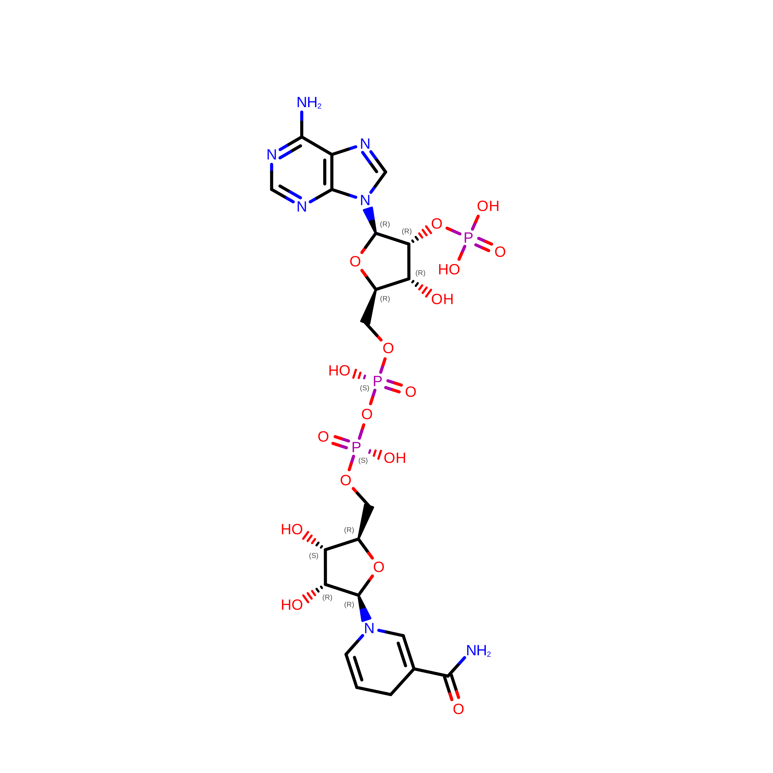
NDP

Query on NDP


Download:Ideal Coordinates CCD File
B [auth A]NADPH DIHYDRO-NICOTINAMIDE-ADENINE-DINUCLEOTIDE PHOSPHATE
C21 H30 N7 O17 P3
ACFIXJIJDZMPPO-NNYOXOHSSA-N
Q74

Query on Q74


Download:Ideal Coordinates CCD File
C [auth A]2-(3-((4,5,7-trifluorobenzo[d]thiazol-2-yl)methyl)-1H-pyrrolo[2,3-b]pyridin-1-yl)acetic acid
C17 H10 F3 N3 O2 S
IEVFQDJUDLCOQY-UHFFFAOYSA-N
Binding Affinity Annotations 
IDSourceBinding Affinity
Q74 BindingDB:  3G5E IC50: 1.00e+5 (nM) from 1 assay(s)

Experimental Data & Validation

Experimental Data

  • Method: X-RAY DIFFRACTION
  • Resolution: 1.80 Å
  • R-Value Free:  0.183 (Depositor), 0.182 (DCC) 
  • R-Value Work:  0.144 (Depositor), 0.144 (DCC) 
  • R-Value Observed: 0.146 (Depositor) 
Space Group: P 1 21 1
Unit Cell:
Length ( Å )Angle ( ˚ )
a = 50.016α = 90
b = 67.204β = 92.58
c = 47.861γ = 90
Software Package:
Software NamePurpose
MAR345dtbdata collection
X-PLORmodel building
REFMACrefinement
DENZOdata reduction
SCALEPACKdata scaling
X-PLORphasing

Structure Validation

View Full Validation Report


Ligand Structure Quality Assessment 


Entry History 

Deposition Data

Revision History  (Full details and data files)

  • Version 1.0: 2009-03-24
    Type: Initial release
  • Version 1.1: 2011-07-13
    Changes: Version format compliance
  • Version 1.2: 2024-03-20
    Changes: Data collection, Database references, Derived calculations