3G53

Ligand migration and cavities within scapharca dimeric hemoglobin: wild type with co bound to heme and chloropropyl benzene bound to the XE4 cavity


Experimental Data Snapshot

  • Method: X-RAY DIFFRACTION
  • Resolution: 1.64 Å
  • R-Value Free: 0.204 
  • R-Value Work: 0.189 
  • R-Value Observed: 0.189 

wwPDB Validation   3D Report Full Report


Ligand Structure Quality Assessment 


This is version 1.3 of the entry. See complete history


Literature

Ligand migration and cavities within Scapharca Dimeric HbI: studies by time-resolved crystallo-graphy, Xe binding, and computational analysis.

Knapp, J.E.Pahl, R.Cohen, J.Nichols, J.C.Schulten, K.Gibson, Q.H.Srajer, V.Royer, W.E.

(2009) Structure 17: 1494-1504

  • DOI: https://doi.org/10.1016/j.str.2009.09.004
  • Primary Citation of Related Structures:  
    3G46, 3G4Q, 3G4R, 3G4U, 3G4V, 3G4W, 3G4Y, 3G52, 3G53

  • PubMed Abstract: 

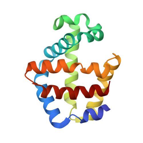
    As in many other hemoglobins, no direct route for migration of ligands between solvent and active site is evident from crystal structures of Scapharca inaequivalvis dimeric HbI. Xenon (Xe) and organic halide binding experiments, along with computational analysis presented here, reveal protein cavities as potential ligand migration routes. Time-resolved crystallographic experiments show that photodissociated carbon monoxide (CO) docks within 5 ns at the distal pocket B site and at more remote Xe4 and Xe2 cavities. CO rebinding is not affected by the presence of dichloroethane within the major Xe4 protein cavity, demonstrating that this cavity is not on the major exit pathway. The crystal lattice has a substantial influence on ligand migration, suggesting that significant conformational rearrangements may be required for ligand exit. Taken together, these results are consistent with a distal histidine gate as one important ligand entry and exit route, despite its participation in the dimeric interface.


  • Organizational Affiliation

    Department of Biochemistry and Molecular Pharmacology, The University of Massachusetts Medical School, Worcester, MA 01605, USA.


Macromolecules
Find similar proteins by:  (by identity cutoff)  |  3D Structure
Entity ID: 1
MoleculeChains Sequence LengthOrganismDetailsImage
Globin-1
A, B
146Anadara inaequivalvisMutation(s): 0 
UniProt
Find proteins for P02213 (Anadara inaequivalvis)
Explore P02213 
Go to UniProtKB:  P02213
Entity Groups  
Sequence Clusters30% Identity50% Identity70% Identity90% Identity95% Identity100% Identity
UniProt GroupP02213
Sequence Annotations
Expand
  • Reference Sequence
Experimental Data & Validation

Experimental Data

  • Method: X-RAY DIFFRACTION
  • Resolution: 1.64 Å
  • R-Value Free: 0.204 
  • R-Value Work: 0.189 
  • R-Value Observed: 0.189 
  • Space Group: C 1 2 1
Unit Cell:
Length ( Å )Angle ( ˚ )
a = 93.19α = 90
b = 43.87β = 121.88
c = 83.35γ = 90
Software Package:
Software NamePurpose
MAR345dtbdata collection
CNSrefinement
DENZOdata reduction
SCALEPACKdata scaling
CNSphasing

Structure Validation

View Full Validation Report


Ligand Structure Quality Assessment 


Entry History 

Deposition Data

Revision History  (Full details and data files)

  • Version 1.0: 2009-12-01
    Type: Initial release
  • Version 1.1: 2011-07-13
    Changes: Version format compliance
  • Version 1.2: 2020-06-24
    Changes: Database references, Source and taxonomy, Structure summary
  • Version 1.3: 2023-09-06
    Changes: Data collection, Database references, Derived calculations, Refinement description