3G26

Crystal structure of the C-terminal domain of the Rous Sarcoma Virus capsid protein: Mutant A184C


Experimental Data Snapshot

  • Method: X-RAY DIFFRACTION
  • Resolution: 1.55 Å
  • R-Value Free: 0.214 
  • R-Value Work: 0.187 
  • R-Value Observed: 0.188 

wwPDB Validation   3D Report Full Report


This is version 1.4 of the entry. See complete history


Literature

Proton-linked dimerization of a retroviral capsid protein initiates capsid assembly

Bailey, G.D.Hyun, J.K.Mitra, A.K.Kingston, R.L.

(2009) Structure 17: 737-748

  • DOI: https://doi.org/10.1016/j.str.2009.03.010
  • Primary Citation of Related Structures:  
    3G0V, 3G1G, 3G1I, 3G21, 3G26, 3G28, 3G29

  • PubMed Abstract: 

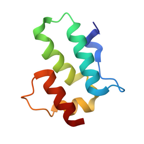
    In mature retroviral particles, the capsid protein (CA) forms a shell encasing the viral replication complex. Human immunodeficiency virus (HIV) CA dimerizes in solution, through its C-terminal domain (CTD), and this interaction is important for capsid assembly. In contrast, other retroviral capsid proteins, including that of Rous sarcoma virus (RSV), do not dimerize with measurable affinity. Here we show, using X-ray crystallography and other biophysical methods, that acidification causes RSV CA to dimerize in a fashion analogous to HIV CA, and that this drives capsid assembly in vitro. A pair of aspartic acid residues, located within the CTD dimer interface, explains why dimerization is linked to proton binding. Our results show that despite overarching structural similarities, the intermolecular forces responsible for forming and stabilizing the retroviral capsid differ markedly across retroviral genera. Our data further suggest that proton binding may regulate RSV capsid assembly, or modulate stability of the assembled capsid.


  • Organizational Affiliation

    School of Biological Sciences, University of Auckland, Auckland, New Zealand.


Macromolecules
Find similar proteins by:  (by identity cutoff)  |  3D Structure
Entity ID: 1
MoleculeChains Sequence LengthOrganismDetailsImage
Gag polyprotein77Rous sarcoma virus - Prague CMutation(s): 1 
Gene Names: gag
EC: 3.4.23
UniProt
Find proteins for P03322 (Rous sarcoma virus subgroup C (strain Prague))
Explore P03322 
Go to UniProtKB:  P03322
Entity Groups  
Sequence Clusters30% Identity50% Identity70% Identity90% Identity95% Identity100% Identity
UniProt GroupP03322
Sequence Annotations
Expand
  • Reference Sequence
Small Molecules
Ligands 1 Unique
IDChains Name / Formula / InChI Key2D Diagram3D Interactions
MLA
Query on MLA

Download Ideal Coordinates CCD File 
B [auth A]MALONIC ACID
C3 H4 O4
OFOBLEOULBTSOW-UHFFFAOYSA-N
Experimental Data & Validation

Experimental Data

  • Method: X-RAY DIFFRACTION
  • Resolution: 1.55 Å
  • R-Value Free: 0.214 
  • R-Value Work: 0.187 
  • R-Value Observed: 0.188 
  • Space Group: P 32 2 1
Unit Cell:
Length ( Å )Angle ( ˚ )
a = 32.243α = 90
b = 32.243β = 90
c = 113.993γ = 120
Software Package:
Software NamePurpose
Blu-Icedata collection
PHASERphasing
REFMACrefinement
HKL-2000data reduction
HKL-2000data scaling

Structure Validation

View Full Validation Report


Entry History 

Deposition Data

Revision History  (Full details and data files)

  • Version 1.0: 2009-06-02
    Type: Initial release
  • Version 1.1: 2011-07-13
    Changes: Version format compliance
  • Version 1.2: 2015-04-29
    Changes: Non-polymer description
  • Version 1.3: 2021-11-10
    Changes: Database references, Derived calculations
  • Version 1.4: 2023-11-01
    Changes: Data collection, Refinement description