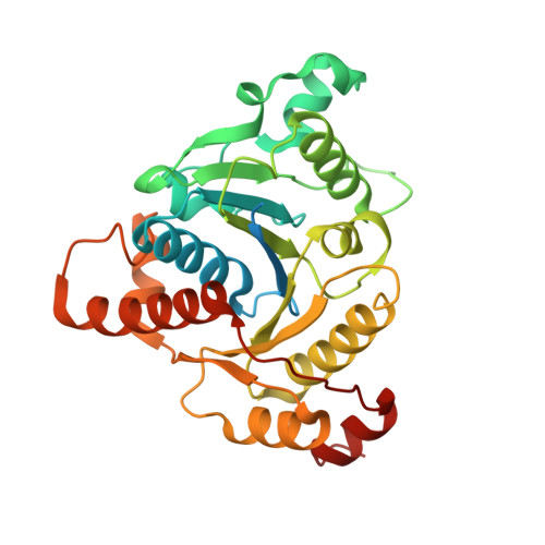
3FWY

Crystal structure of the L protein of Rhodobacter sphaeroides light-independent protochlorophyllide reductase (BchL) with MgADP bound: a homologue of the nitrogenase Fe protein


Experimental Data Snapshot

  • Method: X-RAY DIFFRACTION
  • Resolution: 1.63 Å
  • R-Value Free: 0.199 
  • R-Value Work: 0.174 
  • R-Value Observed: 0.175 

wwPDB Validation   3D Report Full Report


Ligand Structure Quality Assessment 


This is version 1.3 of the entry. See complete history


Literature

Crystal Structure of the L Protein of Rhodobacter sphaeroides Light-Independent Protochlorophyllide Reductase with MgADP Bound: A Homologue of the Nitrogenase Fe Protein.

Sarma, R.Barney, B.M.Hamilton, T.L.Jones, A.Seefeldt, L.C.Peters, J.W.

(2008) Biochemistry 47: 13004-13015

  • DOI: https://doi.org/10.1021/bi801058r
  • Primary Citation of Related Structures:  
    3FWY

  • PubMed Abstract: 
  • The L protein (BchL) of the dark-operative protochlorophyllide reductase (DPOR) from Rhodobacter sphaeroides has been purified from an Azotobacter vinelandii expression system; its interaction with nucleotides has been examined, and the X-ray structure of the protein has been determined with bound MgADP to 1 ...

    The L protein (BchL) of the dark-operative protochlorophyllide reductase (DPOR) from Rhodobacter sphaeroides has been purified from an Azotobacter vinelandii expression system; its interaction with nucleotides has been examined, and the X-ray structure of the protein has been determined with bound MgADP to 1.6 A resolution. DPOR catalyzes the reduction of protochlorophyllide to chlorophyllide, a reaction critical to the biosynthesis of bacteriochlorophylls. The DPOR holoenzyme is comprised of two component proteins, the dimeric BchL protein and the heterotetrameric BchN/BchB protein. The DPOR component proteins share significant overall similarities with the nitrogenase Fe protein (NifH) and the MoFe (NifDK) protein, the enzyme system responsible for reduction of dinitrogen to ammonia. Here, BchL was expressed in A. vinelandii and purified to homogeneity using an engineered polyhistidine tag. The purified, recombinant BchL was found to contain 3.6 mol of Fe/mol of BchL homodimer, consistent with the presence of a [4Fe-4S] cluster and analogous to the [4Fe-4S] cluster present in the Fe protein. The MgATP- and MgADP-induced conformational changes in BchL were examined by an Fe chelation assay and found to be distinctly different from the nucleotide-stimulated Fe release observed for the Fe protein. The recombinant BchL was crystallized with bound MgADP, and the structure was determined to 1.6 A resolution. BchL is found to share overall structural similarity with the nitrogenase Fe protein, including the subunit bridging [4Fe-4S] cluster and nucleotide binding sites. Despite the high level of structural similarity, however, BchL is found to be incapable of substituting for the Fe protein in a nitrogenase substrate reduction assay. The newly determined structure of BchL and its comparison to its close homologue, the nitrogenase Fe protein, provide the basis for understanding how these highly related proteins can discriminate between their respective functions in microbial systems where each must function simultaneously.


    Organizational Affiliation

    Department of Chemistry and Biochemistry and Astrobiology Biogeocatalysis Research Center, Montana State University, Bozeman, Montana 59717, USA.


Macromolecules
Find similar proteins by:  (by identity cutoff)  |  3D Structure
Entity ID: 1
MoleculeChainsSequence LengthOrganismDetailsImage
Light-independent protochlorophyllide reductase iron-sulfur ATP-binding protein
A, B
314Cereibacter sphaeroides 2.4.1Mutation(s): 0 
Gene Names: bchLRHOS4_18930RSP_0288
EC: 1.18 (PDB Primary Data), 1.3.7.7 (UniProt)
UniProt
Find proteins for Q9RFD6 (Cereibacter sphaeroides (strain ATCC 17023 / DSM 158 / JCM 6121 / CCUG 31486 / LMG 2827 / NBRC 12203 / NCIMB 8253 / ATH 2.4.1.))
Explore Q9RFD6 
Go to UniProtKB:  Q9RFD6
Entity Groups  
Sequence Clusters30% Identity50% Identity70% Identity90% Identity95% Identity100% Identity
UniProt GroupQ9RFD6
Protein Feature View
Expand
  • Reference Sequence
Small Molecules
Ligands 3 Unique
IDChainsName / Formula / InChI Key2D Diagram3D Interactions
ADP
Query on ADP

Download Ideal Coordinates CCD File 
D [auth A],
G [auth B]
ADENOSINE-5'-DIPHOSPHATE
C10 H15 N5 O10 P2
XTWYTFMLZFPYCI-KQYNXXCUSA-N
 Ligand Interaction
SF4
Query on SF4

Download Ideal Coordinates CCD File 
F [auth B]IRON/SULFUR CLUSTER
Fe4 S4
LJBDFODJNLIPKO-UHFFFAOYSA-N
 Ligand Interaction
MG
Query on MG

Download Ideal Coordinates CCD File 
C [auth A],
E [auth B]
MAGNESIUM ION
Mg
JLVVSXFLKOJNIY-UHFFFAOYSA-N
 Ligand Interaction
Experimental Data & Validation

Experimental Data

  • Method: X-RAY DIFFRACTION
  • Resolution: 1.63 Å
  • R-Value Free: 0.199 
  • R-Value Work: 0.174 
  • R-Value Observed: 0.175 
  • Space Group: P 21 21 21
Unit Cell:
Length ( Å )Angle ( ˚ )
a = 56.729α = 90
b = 86.622β = 90
c = 117.169γ = 90
Software Package:
Software NamePurpose
HKL-2000data collection
PHASERphasing
REFMACrefinement
HKL-2000data reduction
HKL-2000data scaling

Structure Validation

View Full Validation Report


Ligand Structure Quality Assessment 


Entry History 

Deposition Data

Revision History  (Full details and data files)

  • Version 1.0: 2009-03-24
    Type: Initial release
  • Version 1.1: 2011-07-13
    Changes: Advisory, Version format compliance
  • Version 1.2: 2017-11-01
    Changes: Refinement description
  • Version 1.3: 2023-09-06
    Changes: Data collection, Database references, Derived calculations, Refinement description