3FWP

X-ray structure of uridine nucleoside phosphorylease from Salmonella typhimurium complexed with phosphate and its inhibitor 2,2'-anhydrouridine at 1.86 A resolution


Experimental Data Snapshot

  • Method: X-RAY DIFFRACTION
  • Resolution: 1.86 Å
  • R-Value Free: 0.206 
  • R-Value Work: 0.176 
  • R-Value Observed: 0.178 

wwPDB Validation   3D Report Full Report


Ligand Structure Quality Assessment 


This is version 1.3 of the entry. See complete history


Literature

The X-ray structure of Salmonella typhimurium uridine nucleoside phosphorylase complexed with 2,2'-anhydrouridine, phosphate and potassium ions at 1.86 A resolution.

Lashkov, A.A.Zhukhlistova, N.E.Gabdoulkhakov, A.H.Shtil, A.A.Efremov, R.G.Betzel, C.Mikhailov, A.M.

(2010) Acta Crystallogr D Biol Crystallogr 66: 51-60

  • DOI: https://doi.org/10.1107/S0907444909044175
  • Primary Citation of Related Structures:  
    3FWP

  • PubMed Abstract: 

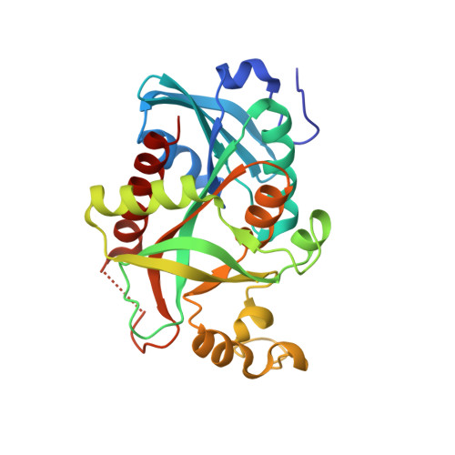
    Uridine nucleoside phosphorylase is an important drug target for the development of anti-infective and antitumour agents. The X-ray crystal structure of Salmonella typhimurium uridine nucleoside phosphorylase (StUPh) complexed with its inhibitor 2,2'-anhydrouridine, phosphate and potassium ions has been solved and refined at 1.86 A resolution (R(cryst) = 17.6%, R(free) = 20.6%). The complex of human uridine phosphorylase I (HUPhI) with 2,2'-anhydrouridine was modelled using a computational approach. The model allowed the identification of atomic groups in 2,2'-anhydrouridine that might improve the interaction of future inhibitors with StUPh and HUPhI.


  • Organizational Affiliation

    A. V. Shubnikov Institute of Crystallography, Russian Academy of Sciences, 59 Leninsky Prospect, 119333 Moscow, Russia.


Macromolecules
Find similar proteins by:  (by identity cutoff)  |  3D Structure
Entity ID: 1
MoleculeChains Sequence LengthOrganismDetailsImage
Uridine phosphorylase
A, B, C, D, E
A, B, C, D, E, F
253Salmonella enterica subsp. enterica serovar TyphimuriumMutation(s): 0 
Gene Names: STM3968STMD1.21udp
EC: 2.4.2.3
UniProt
Find proteins for P0A1F6 (Salmonella typhimurium (strain LT2 / SGSC1412 / ATCC 700720))
Explore P0A1F6 
Go to UniProtKB:  P0A1F6
Entity Groups  
Sequence Clusters30% Identity50% Identity70% Identity90% Identity95% Identity100% Identity
UniProt GroupP0A1F6
Sequence Annotations
Expand
  • Reference Sequence
Small Molecules
Ligands 3 Unique
IDChains Name / Formula / InChI Key2D Diagram3D Interactions
ANU
Query on ANU

Download Ideal Coordinates CCD File 
I [auth B],
L [auth D],
O [auth F]
2,2'-Anhydro-(1-beta-D-arabinofuranosyl)uracil
C9 H10 N2 O5
UUGITDASWNOAGG-CCXZUQQUSA-N
PO4
Query on PO4

Download Ideal Coordinates CCD File 
G [auth B],
K [auth D],
N [auth F]
PHOSPHATE ION
O4 P
NBIIXXVUZAFLBC-UHFFFAOYSA-K
K
Query on K

Download Ideal Coordinates CCD File 
H [auth B],
J [auth C],
M [auth F]
POTASSIUM ION
K
NPYPAHLBTDXSSS-UHFFFAOYSA-N
Experimental Data & Validation

Experimental Data

  • Method: X-RAY DIFFRACTION
  • Resolution: 1.86 Å
  • R-Value Free: 0.206 
  • R-Value Work: 0.176 
  • R-Value Observed: 0.178 
  • Space Group: P 21 21 21
Unit Cell:
Length ( Å )Angle ( ˚ )
a = 88.79α = 90
b = 124.07β = 90
c = 134.1γ = 90
Software Package:
Software NamePurpose
DENZOdata reduction
MOLREPphasing
REFMACrefinement
XDSdata reduction
XSCALEdata scaling

Structure Validation

View Full Validation Report


Ligand Structure Quality Assessment 


Entry History 

Deposition Data

Revision History  (Full details and data files)

  • Version 1.0: 2010-01-19
    Type: Initial release
  • Version 1.1: 2011-07-13
    Changes: Version format compliance
  • Version 1.2: 2017-10-18
    Changes: Refinement description, Structure summary
  • Version 1.3: 2023-09-06
    Changes: Data collection, Database references, Derived calculations, Refinement description