


Structural roles of biocovalent flaninylation in berberine bridge enzyme
Winkler, A., Motz, K., Riedl, S., Puhl, M., Macheroux, P., Gruber, K.To be published.
Experimental Data Snapshot
Starting Model: experimental
View more details
Entity ID: 1 | |||||
|---|---|---|---|---|---|
| Molecule | Chains | Sequence Length | Organism | Details | Image |
| Reticuline oxidase | 495 | Eschscholzia californica | Mutation(s): 0 Gene Names: BBE1 EC: 1.21.3.3 | 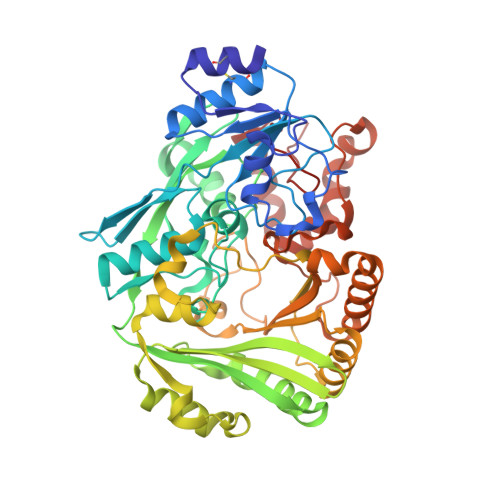 | |
UniProt | |||||
Entity Groups | |||||
| Sequence Clusters | 30% Identity50% Identity70% Identity90% Identity95% Identity100% Identity | ||||
| UniProt Group | P30986 | ||||
Glycosylation | |||||
| Glycosylation Sites: 2 | |||||
Sequence AnnotationsExpand | |||||
Reference Sequence | |||||
| Ligands 4 Unique | |||||
|---|---|---|---|---|---|
| ID | Chains | Name / Formula / InChI Key | 2D Diagram | 3D Interactions | |
| FAD Download:Ideal Coordinates CCD File | C [auth A] | FLAVIN-ADENINE DINUCLEOTIDE C27 H33 N9 O15 P2 VWWQXMAJTJZDQX-UYBVJOGSSA-N |  | ||
| SLX Download:Ideal Coordinates CCD File | D [auth A] | (13aS)-3,10-dimethoxy-5,8,13,13a-tetrahydro-6H-isoquino[3,2-a]isoquinoline-2,9-diol C19 H21 N O4 KNWVMRVOBAFFMH-HNNXBMFYSA-N |  | ||
| NAG Download:Ideal Coordinates CCD File | E [auth A] | 2-acetamido-2-deoxy-beta-D-glucopyranose C8 H15 N O6 OVRNDRQMDRJTHS-FMDGEEDCSA-N |  | ||
| MG Download:Ideal Coordinates CCD File | F [auth A] | MAGNESIUM ION Mg JLVVSXFLKOJNIY-UHFFFAOYSA-N |  | ||
| Length ( Å ) | Angle ( ˚ ) |
|---|---|
| a = 68.72 | α = 90 |
| b = 68.72 | β = 90 |
| c = 247.17 | γ = 90 |
| Software Name | Purpose |
|---|---|
| PHENIX | refinement |
| PDB_EXTRACT | data extraction |
| MAR345dtb | data collection |
| XDS | data reduction |
| XSCALE | data scaling |
| PHASER | phasing |


