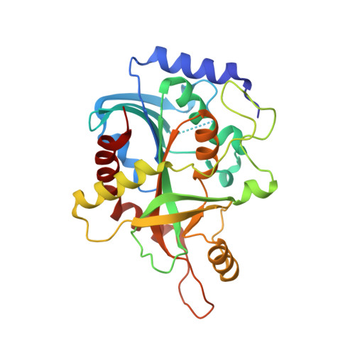
1.45 A resolution crystal structure of recombinant PNP in complex with a pM multisubstrate analogue inhibitor bearing one feature of the postulated transition state.
Chojnowski, G., Breer, K., Narczyk, M., Wielgus-Kutrowska, B., Czapinska, H., Hashimoto, M., Hikishima, S., Yokomatsu, T., Bochtler, M., Girstun, A., Staron, K., Bzowska, A.(2010) Biochem Biophys Res Commun 391: 703-708
- PubMed: 19944078 Search on PubMed
- DOI: https://doi.org/10.1016/j.bbrc.2009.11.124
- Primary Citation Related Structures:
3FUC - PubMed Abstract:
Low molecular mass purine nucleoside phosphorylases (PNPs, E.C. 2.4.2.1) are homotrimeric enzymes that are tightly inhibited by immucillins. Due to the positive charge on the ribose like part (iminoribitol moiety) and protonation of the N7 atom of the purine ring, immucillins are believed to act as transition state analogues. Over a wide range of concentrations, immucillins bind with strong negative cooperativity to PNPs, so that only every third binding site of the enzyme is occupied (third-of-the-sites binding). 9-(5',5'-difluoro-5'-phosphonopentyl)-9-deazaguanine (DFPP-DG) shares with immucillins the protonation of the N7, but not the positive charge on the ribose like part of the molecule. We have previously shown that DFPP-DG interacts with PNPs with subnanomolar inhibition constant. Here, we report additional biochemical experiments to demonstrate that the inhibitor can be bound with the same K(d) ( approximately 190pM) to all three substrate binding sites of the trimeric PNP, and a crystal structure of PNP in complex with DFPP-DG at 1.45A resolution, the highest resolution published for PNPs so far. The crystals contain the full PNP homotrimer in the asymmetric unit. DFPP-DG molecules are bound in superimposable manner and with full occupancies to all three PNP subunits. Thus the postulated third-of-the-sites binding of immucillins should be rather attribute to the second feature of the transition state, ribooxocarbenium ion character of the ligand or to the coexistence of both features characteristic for the transition state. The DFPP-DG/PNP complex structure confirms the earlier observations, that the loop from Pro57 to Gly66 covering the phosphate-binding site cannot be stabilized by phosphonate analogues. The loop from Glu250 to Gln266 covering the base-binding site is organized by the interactions of Asn243 with the Hoogsteen edge of the purine base of analogues bearing one feature of the postulated transition state (protonated N7 position).
- Department of Biophysics, Institute of Experimental Physics, University of Warsaw, Zwirki i Wigury 93, 02-089 Warsaw, Poland.
Organizational Affiliation: