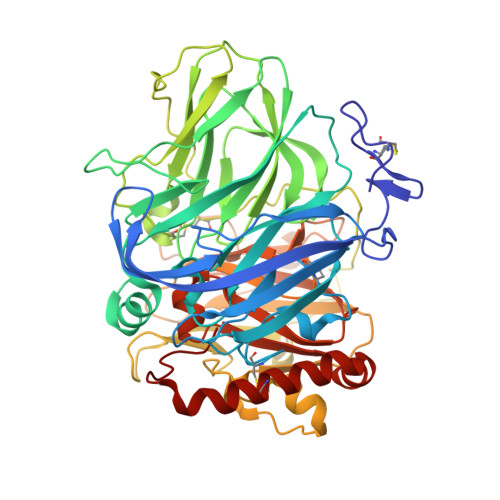
3FU8

Melanocarpus albomyces laccase crystal soaked (10 sec) with 2,6-dimethoxyphenol


Experimental Data Snapshot

  • Method: X-RAY DIFFRACTION
  • Resolution: 1.80 Å
  • R-Value Free: 0.256 
  • R-Value Work: 0.203 
  • R-Value Observed: 0.206 

wwPDB Validation   3D Report Full Report


Ligand Structure Quality Assessment 


This is version 2.0 of the entry. See complete history


Literature

Structure-Function Studies of a Melanocarpus albomyces Laccase Suggest a Pathway for Oxidation of Phenolic Compounds.

Kallio, J.P.Auer, S.Janis, J.Andberg, M.Kruus, K.Rouvinen, J.Koivula, A.Hakulinen, N.

(2009) J Mol Biol 392: 895-909

  • DOI: https://doi.org/10.1016/j.jmb.2009.06.053
  • Primary Citation of Related Structures:  
    3FU7, 3FU8, 3FU9

  • PubMed Abstract: 

    Melanocarpus albomyces laccase crystals were soaked with 2,6-dimethoxyphenol, a common laccase substrate. Three complex structures from different soaking times were solved. Crystal structures revealed the binding of the original substrate and adducts formed by enzymatic oxidation of the substrate. The dimeric oxidation products were identified by mass spectrometry. In the crystals, a 2,6-dimethoxy-p-benzoquinone and a C-O dimer were observed, whereas a C-C dimer was the main product identified by mass spectrometry. Crystal structures demonstrated that the substrate and/or its oxidation products were bound in the pocket formed by residues Ala191, Pro192, Glu235, Leu363, Phe371, Trp373, Phe427, Leu429, Trp507 and His508. Substrate and adducts were hydrogen-bonded to His508, one of the ligands of type 1 copper. Therefore, this surface-exposed histidine most likely has a role in electron transfer by laccases. Based on our mutagenesis studies, the carboxylic acid residue Glu235 at the bottom of the binding site pocket is also crucial in the oxidation of phenolics. Glu235 may be responsible for the abstraction of a proton from the OH group of the substrate and His508 may extract an electron. In addition, crystal structures revealed a secondary binding site formed through weak dimerization in M. albomyces laccase molecules. This binding site most likely exists only in crystals, when the Phe427 residues are packed against each other.


  • Organizational Affiliation

    Department of Chemistry, University of Joensuu, P.O. Box 111, FIN-80101 Joensuu, Finland.


Macromolecules
Find similar proteins by:  (by identity cutoff)  |  3D Structure
Entity ID: 1
MoleculeChains Sequence LengthOrganismDetailsImage
Laccase-1
A, B
559Melanocarpus albomycesMutation(s): 0 
Gene Names: LAC1
EC: 1.10.3.2
UniProt
Find proteins for Q70KY3 (Melanocarpus albomyces)
Explore Q70KY3 
Go to UniProtKB:  Q70KY3
Entity Groups  
Sequence Clusters30% Identity50% Identity70% Identity90% Identity95% Identity100% Identity
UniProt GroupQ70KY3
Glycosylation
Glycosylation Sites: 7
Sequence Annotations
Expand
  • Reference Sequence
Oligosaccharides

Help

Entity ID: 2
MoleculeChains Length2D Diagram Glycosylation3D Interactions
alpha-D-mannopyranose-(1-6)-alpha-D-mannopyranose-(1-4)-2-acetamido-2-deoxy-beta-D-glucopyranose-(1-4)-2-acetamido-2-deoxy-beta-D-glucopyranose
C, H
4N-Glycosylation
Glycosylation Resources
GlyTouCan:  G04854NQ
GlyCosmos:  G04854NQ
GlyGen:  G04854NQ
Entity ID: 3
MoleculeChains Length2D Diagram Glycosylation3D Interactions
2-acetamido-2-deoxy-beta-D-glucopyranose-(1-4)-2-acetamido-2-deoxy-beta-D-glucopyranose
D, F, G, I, K
2N-Glycosylation
Glycosylation Resources
GlyTouCan:  G42666HT
GlyCosmos:  G42666HT
GlyGen:  G42666HT
Entity ID: 4
MoleculeChains Length2D Diagram Glycosylation3D Interactions
alpha-D-mannopyranose-(1-4)-2-acetamido-2-deoxy-beta-D-glucopyranose-(1-4)-2-acetamido-2-deoxy-beta-D-glucopyranose
E, J
3N-Glycosylation
Glycosylation Resources
GlyTouCan:  G62182OO
GlyCosmos:  G62182OO
GlyGen:  G62182OO
Small Molecules
Ligands 6 Unique
IDChains Name / Formula / InChI Key2D Diagram3D Interactions
NAG
Query on NAG

Download Ideal Coordinates CCD File 
BA [auth B],
CA [auth B],
DA [auth B],
Q [auth A],
R [auth A]
2-acetamido-2-deoxy-beta-D-glucopyranose
C8 H15 N O6
OVRNDRQMDRJTHS-FMDGEEDCSA-N
3DM
Query on 3DM

Download Ideal Coordinates CCD File 
FA [auth B],
U [auth A]
2,6-dimethoxyphenol
C8 H10 O3
KLIDCXVFHGNTTM-UHFFFAOYSA-N
SO4
Query on SO4

Download Ideal Coordinates CCD File 
EA [auth B],
S [auth A],
T [auth A]
SULFATE ION
O4 S
QAOWNCQODCNURD-UHFFFAOYSA-L
GOL
Query on GOL

Download Ideal Coordinates CCD File 
GA [auth B],
V [auth A]
GLYCEROL
C3 H8 O3
PEDCQBHIVMGVHV-UHFFFAOYSA-N
CU
Query on CU

Download Ideal Coordinates CCD File 
L [auth A]
M [auth A]
N [auth A]
O [auth A]
W [auth B]
L [auth A],
M [auth A],
N [auth A],
O [auth A],
W [auth B],
X [auth B],
Y [auth B],
Z [auth B]
COPPER (II) ION
Cu
JPVYNHNXODAKFH-UHFFFAOYSA-N
CL
Query on CL

Download Ideal Coordinates CCD File 
AA [auth B],
P [auth A]
CHLORIDE ION
Cl
VEXZGXHMUGYJMC-UHFFFAOYSA-M
Modified Residues  1 Unique
IDChains TypeFormula2D DiagramParent
OHI
Query on OHI
A, B
L-PEPTIDE LINKINGC6 H7 N3 O3HIS
Experimental Data & Validation

Experimental Data

  • Method: X-RAY DIFFRACTION
  • Resolution: 1.80 Å
  • R-Value Free: 0.256 
  • R-Value Work: 0.203 
  • R-Value Observed: 0.206 
  • Space Group: C 1 2 1
Unit Cell:
Length ( Å )Angle ( ˚ )
a = 174.71α = 90
b = 62.37β = 96.36
c = 125.14γ = 90
Software Package:
Software NamePurpose
MOLREPphasing
REFMACrefinement
XDSdata reduction
XSCALEdata scaling

Structure Validation

View Full Validation Report


Ligand Structure Quality Assessment 


Entry History 

Deposition Data

Revision History  (Full details and data files)

  • Version 1.0: 2009-09-22
    Type: Initial release
  • Version 1.1: 2011-07-13
    Changes: Non-polymer description, Version format compliance
  • Version 2.0: 2020-07-29
    Type: Remediation
    Reason: Carbohydrate remediation
    Changes: Advisory, Atomic model, Data collection, Derived calculations, Structure summary