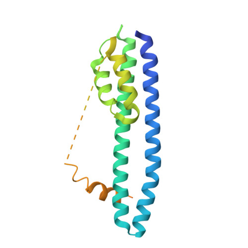
3FRT

The structure of human CHMP3 (residues 8 - 222).


Experimental Data Snapshot

  • Method: X-RAY DIFFRACTION
  • Resolution: 4.00 Å
  • R-Value Free: 0.436 
  • R-Value Work: 0.383 
  • R-Value Observed: 0.388 

Starting Model: experimental
View more details

wwPDB Validation   3D Report Full Report


This is version 1.3 of the entry. See complete history


Literature

Structural basis for ESCRT-III protein autoinhibition.

Bajorek, M.Schubert, H.L.McCullough, J.Langelier, C.Eckert, D.M.Stubblefield, W.M.Uter, N.T.Myszka, D.G.Hill, C.P.Sundquist, W.I.

(2009) Nat Struct Mol Biol 16: 754-762

  • DOI: https://doi.org/10.1038/nsmb.1621
  • Primary Citation of Related Structures:  
    3FRR, 3FRS, 3FRT, 3FRV

  • PubMed Abstract: 

    Endosomal sorting complexes required for transport-III (ESCRT-III) subunits cycle between two states: soluble monomers and higher-order assemblies that bind and remodel membranes during endosomal vesicle formation, midbody abscission and enveloped virus budding. Here we show that the N-terminal core domains of increased sodium tolerance-1 (IST1) and charged multivesicular body protein-3 (CHMP3) form equivalent four-helix bundles, revealing that IST1 is a previously unrecognized ESCRT-III family member. IST1 and its ESCRT-III binding partner, CHMP1B, both form higher-order helical structures in vitro, and IST1-CHMP1 interactions are required for abscission. The IST1 and CHMP3 structures also reveal that equivalent downstream alpha5 helices can fold back against the core domains. Mutations within the CHMP3 core-alpha5 interface stimulate the protein's in vitro assembly and HIV-inhibition activities, indicating that dissociation of the autoinhibitory alpha5 helix from the core activates ESCRT-III proteins for assembly at membranes.


  • Organizational Affiliation

    Department of Biochemistry, University of Utah, Salt Lake City, Utah, USA.


Macromolecules
Find similar proteins by:  (by identity cutoff)  |  3D Structure
Entity ID: 1
MoleculeChains Sequence LengthOrganismDetailsImage
Charged multivesicular body protein 3
A, B
218Homo sapiensMutation(s): 0 
Gene Names: CGI-149CHMP3NEDFVPS24
UniProt & NIH Common Fund Data Resources
Find proteins for Q9Y3E7 (Homo sapiens)
Explore Q9Y3E7 
Go to UniProtKB:  Q9Y3E7
PHAROS:  Q9Y3E7
GTEx:  ENSG00000115561 
Entity Groups  
Sequence Clusters30% Identity50% Identity70% Identity90% Identity95% Identity100% Identity
UniProt GroupQ9Y3E7
Sequence Annotations
Expand
  • Reference Sequence
Experimental Data & Validation

Experimental Data

  • Method: X-RAY DIFFRACTION
  • Resolution: 4.00 Å
  • R-Value Free: 0.436 
  • R-Value Work: 0.383 
  • R-Value Observed: 0.388 
  • Space Group: P 1 21 1
Unit Cell:
Length ( Å )Angle ( ˚ )
a = 36.472α = 90
b = 131.514β = 108.06
c = 48.508γ = 90
Software Package:
Software NamePurpose
MAR345dtbdata collection
PHASERphasing
HKL-2000data reduction
HKL-2000data scaling

Structure Validation

View Full Validation Report


Entry History 

Deposition Data

Revision History  (Full details and data files)

  • Version 1.0: 2009-06-30
    Type: Initial release
  • Version 1.1: 2011-07-13
    Changes: Version format compliance
  • Version 1.2: 2017-11-01
    Changes: Refinement description
  • Version 1.3: 2023-09-06
    Changes: Data collection, Database references, Refinement description