3FOQ

Crystal structure of N-acetylglucosamine-1-phosphate uridyltransferase (GlmU) from Mycobacterium tuberculosis in a cubic space group.


Experimental Data Snapshot

  • Method: X-RAY DIFFRACTION
  • Resolution: 3.41 Å
  • R-Value Free: 0.321 
  • R-Value Work: 0.285 
  • R-Value Observed: 0.287 

wwPDB Validation   3D Report Full Report


This is version 1.2 of the entry. See complete history


Literature

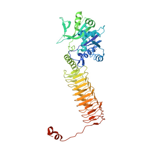
Structure of N-acetylglucosamine-1-phosphate uridyltransferase (GlmU) from Mycobacterium tuberculosis in a cubic space group.

Verma, S.K.Jaiswal, M.Kumar, N.Parikh, A.Nandicoori, V.K.Prakash, B.

(2009) Acta Crystallogr Sect F Struct Biol Cryst Commun 65: 435-439

  • DOI: https://doi.org/10.1107/S1744309109010252
  • Primary Citation of Related Structures:  
    3FOQ

  • PubMed Abstract: 

    GlmU is a bifunctional enzyme that catalyzes the final two steps in the biosynthesis of UDP-GlcNAc. Crystals of GlmU from Mycobacterium tuberculosis obtained using ammonium sulfate as a precipitant diffracted poorly (to 3.4 A resolution) and displayed an unusually high solvent content (>80%) with sparse crystal packing that resulted in large solvent channels. With one molecule per asymmetric unit, the monomers from three neighbouring asymmetric units related by the crystal threefold formed a biological trimer. Although this is the first report of the structure of GlmU determined in a cubic crystal form, the trimeric arrangement here is similar to that observed for other GlmU structures determined in hexagonal (H3, H32, P6(3)22) space groups.


  • Organizational Affiliation

    Department of Biological Sciences and Bioengineering, Indian Institute of Technology, Kanpur, India.


Macromolecules
Find similar proteins by:  (by identity cutoff)  |  3D Structure
Entity ID: 1
MoleculeChains Sequence LengthOrganismDetailsImage
Bifunctional protein glmU503Mycobacterium tuberculosisMutation(s): 0 
Gene Names: glmUMRA_1026MT1046Rv1018c
EC: 2.7.7.23 (PDB Primary Data), 2.3.1.157 (PDB Primary Data)
UniProt
Find proteins for P9WMN3 (Mycobacterium tuberculosis (strain ATCC 25618 / H37Rv))
Explore P9WMN3 
Go to UniProtKB:  P9WMN3
Entity Groups  
Sequence Clusters30% Identity50% Identity70% Identity90% Identity95% Identity100% Identity
UniProt GroupP9WMN3
Sequence Annotations
Expand
  • Reference Sequence
Experimental Data & Validation

Experimental Data

  • Method: X-RAY DIFFRACTION
  • Resolution: 3.41 Å
  • R-Value Free: 0.321 
  • R-Value Work: 0.285 
  • R-Value Observed: 0.287 
  • Space Group: I 4 3 2
Unit Cell:
Length ( Å )Angle ( ˚ )
a = 285.7α = 90
b = 285.7β = 90
c = 285.7γ = 90
Software Package:
Software NamePurpose
XSCALEdata scaling
PHASERphasing
REFMACrefinement
PDB_EXTRACTdata extraction
MAR345dtbdata collection

Structure Validation

View Full Validation Report


Entry History 

Deposition Data

Revision History  (Full details and data files)

  • Version 1.0: 2009-06-09
    Type: Initial release
  • Version 1.1: 2011-07-13
    Changes: Version format compliance
  • Version 1.2: 2024-03-20
    Changes: Data collection, Database references, Derived calculations