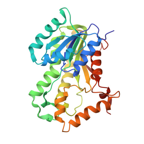
3FNF

Crystal structure of InhA bound to triclosan derivative


Experimental Data Snapshot

  • Method: X-RAY DIFFRACTION
  • Resolution: 2.30 Å
  • R-Value Free: 0.260 
  • R-Value Work: 0.200 
  • R-Value Observed: 0.203 

Starting Model: experimental
View more details

wwPDB Validation   3D Report Full Report


This is version 1.2 of the entry. See complete history


Literature

Triclosan Derivatives: Towards Potent Inhibitors of Drug-Sensitive and Drug-Resistant Mycobacterium tuberculosis.

Freundlich, J.S.Wang, F.Vilcheze, C.Gulten, G.Langley, R.Schiehser, G.A.Jacobus, D.P.Jacobs, W.R.Sacchettini, J.C.

(2009) ChemMedChem 4: 241-248

  • DOI: https://doi.org/10.1002/cmdc.200800261
  • Primary Citation of Related Structures:  
    3FNE, 3FNF, 3FNG, 3FNH

  • PubMed Abstract: 

    Triclosan has been previously shown to inhibit InhA, an essential enoyl acyl carrier protein reductase involved in mycolic acid biosynthesis, the inhibition of which leads to the lysis of Mycobacterium tuberculosis. Using a structure-based drug design approach, a series of 5-substituted triclosan derivatives was developed. Two groups of derivatives with alkyl and aryl substituents, respectively, were identified with dramatically enhanced potency against purified InhA. The most efficacious inhibitor displayed an IC(50) value of 21 nM, which was 50-fold more potent than triclosan. X-ray crystal structures of InhA in complex with four triclosan derivatives revealed the structural basis for the inhibitory activity. Six selected triclosan derivatives were tested against isoniazid-sensitive and resistant strains of M. tuberculosis. Among those, the best inhibitor had an MIC value of 4.7 microg mL(-1) (13 microM), which represents a tenfold improvement over the bacteriocidal activity of triclosan. A subset of these triclosan analogues was more potent than isoniazid against two isoniazid-resistant M. tuberculosis strains, demonstrating the significant potential for structure-based design in the development of next generation antitubercular drugs.


  • Organizational Affiliation

    Texas A&M University, Department of Biochemistry and Biophysics, College Station, 77843-2128, USA.


Macromolecules
Find similar proteins by:  (by identity cutoff)  |  3D Structure
Entity ID: 1
MoleculeChains Sequence LengthOrganismDetailsImage
Enoyl-[acyl-carrier-protein] reductase [NADH]
A, B, C, D
269Mycobacterium tuberculosisMutation(s): 0 
Gene Names: inhARv1484MT1531MTCY277.05
EC: 1.3.1.9
UniProt
Find proteins for P9WGR1 (Mycobacterium tuberculosis (strain ATCC 25618 / H37Rv))
Explore P9WGR1 
Go to UniProtKB:  P9WGR1
Entity Groups  
Sequence Clusters30% Identity50% Identity70% Identity90% Identity95% Identity100% Identity
UniProt GroupP9WGR1
Sequence Annotations
Expand
  • Reference Sequence
Experimental Data & Validation

Experimental Data

  • Method: X-RAY DIFFRACTION
  • Resolution: 2.30 Å
  • R-Value Free: 0.260 
  • R-Value Work: 0.200 
  • R-Value Observed: 0.203 
  • Space Group: C 1 2 1
Unit Cell:
Length ( Å )Angle ( ˚ )
a = 125.651α = 90
b = 92.377β = 106.51
c = 102.406γ = 90
Software Package:
Software NamePurpose
HKL-2000data collection
MOLREPphasing
REFMACrefinement
HKL-2000data reduction
HKL-2000data scaling

Structure Validation

View Full Validation Report


Entry History 

Deposition Data

  • Released Date: 2009-01-20 
  • Deposition Author(s): Wang, F.

Revision History  (Full details and data files)

  • Version 1.0: 2009-01-20
    Type: Initial release
  • Version 1.1: 2011-07-13
    Changes: Version format compliance
  • Version 1.2: 2023-09-06
    Changes: Data collection, Database references, Derived calculations, Refinement description