3FJT | pdb_00003fjt

Crystal structure of a human Fc fragment engineered for extended serum half-life


Experimental Data Snapshot

  • Method: X-RAY DIFFRACTION
  • Resolution: 2.50 Å
  • R-Value Free: 
    0.291 (Depositor), 0.268 (DCC) 
  • R-Value Work: 
    0.227 (Depositor), 0.257 (DCC) 
  • R-Value Observed: 
    0.230 (Depositor) 

wwPDB Validation 3D Report Full Report

Validation slider image for 3FJT

This is version 2.2 of the entry. See complete history

Literature

Structural characterization of a human Fc fragment engineered for extended serum half-life.

Oganesyan, V.Damschroder, M.M.Woods, R.M.Cook, K.E.Wu, H.Dall'acqua, W.F.

(2009) Mol Immunol 46: 1750-1755

  • DOI: https://doi.org/10.1016/j.molimm.2009.01.026
  • Primary Citation Related Structures: 
    3FJT

  • PubMed Abstract: 

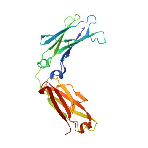
    The first three-dimensional structure of a human Fc fragment genetically engineered for improved pharmacokinetics properties is reported. When introduced into the C(H)2 domain of human immunoglobulin G (IgG) molecules, the triple mutation M252Y/S254T/T256E ('YTE') causes an about 10-fold increase in their binding to the human neonatal Fc receptor (FcRn). This translates into an almost 4-fold increase in the serum half-life of YTE-containing human IgGs in cynomolgus monkeys. A recombinantly produced human Fc/YTE fragment was crystallized and its structure solved at a resolution of 2.5A using molecular replacement. This revealed that Fc/YTE three-dimensional structure is very similar to that of other human Fc fragments in the experimentally visible region spanning residues 236-444. We propose that the enhanced interaction between Fc/YTE and human FcRn is likely mediated by local effects at the substitutions sites. Molecular modeling suggested that potential favorable hydrogen bonds along with an increase in the surface of contact between the two partners may account in part for the corresponding increase in affinity.


  • Organizational Affiliation
    • Department of Antibody Discovery and Protein Engineering, MedImmune, One MedImmune Way, Gaithersburg, MD 20878, USA.

Macromolecule Content 

  • Total Structure Weight: 50.46 kDa 
  • Atom Count: 3,616 
  • Modeled Residue Count: 418 
  • Deposited Residue Count: 418 
  • Unique protein chains: 1

Macromolecules

Find similar proteins by:|  3D Structure
Entity ID: 1
MoleculeChains  Sequence LengthOrganismDetailsImage
Ig gamma-1 chain C region
A, B
209Homo sapiensMutation(s): 3 
Gene Names: IgG1IGHG1
UniProt & NIH Common Fund Data Resources
Find proteins for P01857 (Homo sapiens)
Explore P01857 
Go to UniProtKB:  P01857
PHAROS:  P01857
GTEx:  ENSG00000211896 
Entity Groups
Sequence Clusters30% Identity50% Identity70% Identity90% Identity95% Identity100% Identity
UniProt GroupP01857
Glycosylation
Glycosylation Sites: 1Go to GlyGen: P01857-2
Sequence Annotations
Expand
Reference Sequence

Oligosaccharides

Help  
Entity ID: 2
MoleculeChains Length2D Diagram GlycosylationD Interactions
2-acetamido-2-deoxy-beta-D-glucopyranose-(1-2)-alpha-D-mannopyranose-(1-3)-[2-acetamido-2-deoxy-beta-D-glucopyranose-(1-2)-alpha-D-mannopyranose-(1-6)]beta-D-mannopyranose-(1-4)-2-acetamido-2-deoxy-beta-D-glucopyranose-(1-4)-[alpha-L-fucopyranose-(1-6)]2-acetamido-2-deoxy-beta-D-glucopyranose
C, D
8N-Glycosylation
Glycosylation Resources
GlyTouCan: G80858MF
GlyCosmos: G80858MF
GlyGen: G80858MF

Experimental Data & Validation

Experimental Data

  • Method: X-RAY DIFFRACTION
  • Resolution: 2.50 Å
  • R-Value Free:  0.291 (Depositor), 0.268 (DCC) 
  • R-Value Work:  0.227 (Depositor), 0.257 (DCC) 
  • R-Value Observed: 0.230 (Depositor) 
Space Group: P 21 21 21
Unit Cell:
Length ( Å )Angle ( ˚ )
a = 49.66α = 90
b = 79.605β = 90
c = 145.59γ = 90
Software Package:
Software NamePurpose
DENZOdata reduction
SCALEPACKdata scaling
PHASERphasing
REFMACrefinement
PDB_EXTRACTdata extraction

Structure Validation

View Full Validation Report


Entry History 

Deposition Data

Revision History  (Full details and data files)

  • Version 1.0: 2009-03-24
    Type: Initial release
  • Version 1.1: 2011-07-13
    Changes: Non-polymer description, Version format compliance
  • Version 1.2: 2017-11-01
    Changes: Refinement description
  • Version 2.0: 2020-07-29
    Type: Remediation
    Reason: Carbohydrate remediation
    Changes: Advisory, Atomic model, Data collection, Derived calculations, Structure summary
  • Version 2.1: 2021-10-20
    Changes: Database references, Structure summary
  • Version 2.2: 2024-10-30
    Changes: Data collection, Structure summary