3FGT | pdb_00003fgt

Two chain form of the 66.3 kDa protein from mouse lacking the linker peptide


Experimental Data Snapshot

  • Method: X-RAY DIFFRACTION
  • Resolution: 2.40 Å
  • R-Value Free: 
    0.207 (Depositor), 0.208 (DCC) 
  • R-Value Work: 
    0.166 (Depositor), 0.166 (DCC) 
  • R-Value Observed: 
    0.168 (Depositor) 

Starting Model: experimental
View more details

wwPDB Validation 3D Report Full Report

Validation slider image for 3FGT

Ligand Structure Quality Assessment 


This is version 2.2 of the entry. See complete history

Literature

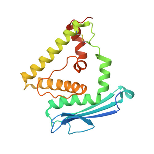
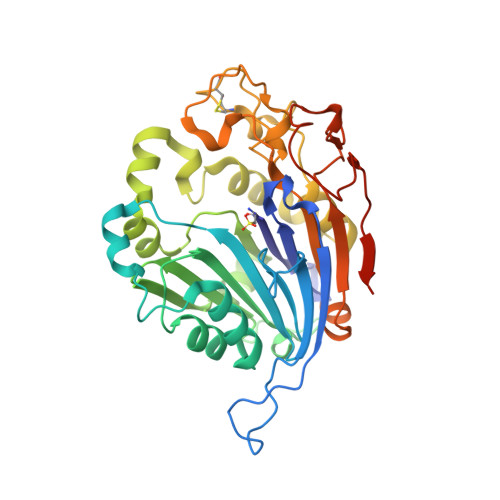
Initial insight into the function of the lysosomal 66.3 kDa protein from mouse by means of X-ray crystallography

Lakomek, K.Dickmanns, A.Kettwig, M.Urlaub, H.Ficner, R.Luebke, T.

(2009) BMC Struct Biol 9: 56-56

  • DOI: https://doi.org/10.1186/1472-6807-9-56
  • Primary Citation Related Structures: 
    3FGR, 3FGT, 3FGW

  • PubMed Abstract: 

    The lysosomal 66.3 kDa protein from mouse is a soluble, mannose 6-phosphate containing protein of so far unknown function. It is synthesized as a glycosylated 75 kDa precursor that undergoes limited proteolysis leading to a 28 kDa N- and a 40 kDa C-terminal fragment. In order to gain insight into the function and the post-translational maturation process of the glycosylated 66.3 kDa protein, three crystal structures were determined that represent different maturation states. These structures demonstrate that the 28 kDa and 40 kDa fragment which have been derived by a proteolytic cleavage remain associated. Mass spectrometric analysis confirmed the subsequent trimming of the C-terminus of the 28 kDa fragment making a large pocket accessible, at the bottom of which the putative active site is located. The crystal structures reveal a significant similarity of the 66.3 kDa protein to several bacterial hydrolases. The core alphabetabetaalpha sandwich fold and a cysteine residue at the N-terminus of the 40 kDa fragment (C249) classify the 66.3 kDa protein as a member of the structurally defined N-terminal nucleophile (Ntn) hydrolase superfamily. Due to the close resemblance of the 66.3 kDa protein to members of the Ntn hydrolase superfamily a hydrolytic activity on substrates containing a non-peptide amide bond seems reasonable. The structural homology which comprises both the overall fold and essential active site residues also implies an autocatalytic maturation process of the lysosomal 66.3 kDa protein. Upon the proteolytic cleavage between S248 and C249, a deep pocket becomes solvent accessible, which harbors the putative active site of the 66.3 kDa protein.


  • Organizational Affiliation
    • Department of Molecular Structural Biology, Institute of Microbiology and Genetics, GZMB, Georg-August University Goettingen, Justus-von-Liebig-Weg 11, D-37077 Goettingen, Germany. klakome@gwdg.de

Macromolecule Content 

  • Total Structure Weight: 65.48 kDa 
  • Atom Count: 4,664 
  • Modeled Residue Count: 524 
  • Deposited Residue Count: 559 
  • Unique protein chains: 2

Macromolecules

Find similar proteins by:|  3D Structure
Entity ID: 1
MoleculeChains  Sequence LengthOrganismDetailsImage
Putative phospholipase B-like 2 28 kDa form202Mus musculusMutation(s): 0 
Gene Names: AAG44101
EC: 3.1.1 (PDB Primary Data), 3.4.11 (UniProt)
UniProt & NIH Common Fund Data Resources
Find proteins for Q3TCN2 (Mus musculus)
Explore Q3TCN2 
Go to UniProtKB:  Q3TCN2
IMPC:  MGI:1919022
Entity Groups
Sequence Clusters30% Identity50% Identity70% Identity90% Identity95% Identity100% Identity
UniProt GroupQ3TCN2
Glycosylation
Glycosylation Sites: 2Go to GlyGen: Q3TCN2-1
Sequence Annotations
Expand
Reference Sequence
Find similar proteins by:|  3D Structure
Entity ID: 2
MoleculeChains  Sequence LengthOrganismDetailsImage
Putative phospholipase B-like 2 40 kDa form357Mus musculusMutation(s): 0 
Gene Names: AAG44101
EC: 3.1.1 (PDB Primary Data), 3.4.11 (UniProt)
UniProt & NIH Common Fund Data Resources
Find proteins for Q3TCN2 (Mus musculus)
Explore Q3TCN2 
Go to UniProtKB:  Q3TCN2
IMPC:  MGI:1919022
Entity Groups
Sequence Clusters30% Identity50% Identity70% Identity90% Identity95% Identity100% Identity
UniProt GroupQ3TCN2
Glycosylation
Glycosylation Sites: 1Go to GlyGen: Q3TCN2-1
Sequence Annotations
Expand
Reference Sequence

Oligosaccharides

Help  
Entity ID: 3
MoleculeChains Length2D Diagram GlycosylationD Interactions
2-acetamido-2-deoxy-beta-D-glucopyranose-(1-4)-2-acetamido-2-deoxy-beta-D-glucopyranose
C
2N-Glycosylation
Glycosylation Resources
GlyTouCan: G42666HT
GlyCosmos: G42666HT
GlyGen: G42666HT

Small Molecules

Ligands 6 Unique
IDChains Name / Formula / InChI Key2D Diagram3D Interactions
7PE

Query on 7PE


Download:Ideal Coordinates CCD File
Q [auth B]2-(2-(2-(2-(2-(2-ETHOXYETHOXY)ETHOXY)ETHOXY)ETHOXY)ETHOXY)ETHANOL
C14 H30 O7
UKXKPKBTMYNOFS-UHFFFAOYSA-N
NAG

Query on NAG


Download:Ideal Coordinates CCD File
D [auth A],
I [auth B]
2-acetamido-2-deoxy-beta-D-glucopyranose
C8 H15 N O6
OVRNDRQMDRJTHS-FMDGEEDCSA-N
PG4

Query on PG4


Download:Ideal Coordinates CCD File
G [auth A],
M [auth B]
TETRAETHYLENE GLYCOL
C8 H18 O5
UWHCKJMYHZGTIT-UHFFFAOYSA-N
GOL

Query on GOL


Download:Ideal Coordinates CCD File
F [auth A],
K [auth B],
L [auth B],
N [auth B],
O [auth B]
GLYCEROL
C3 H8 O3
PEDCQBHIVMGVHV-UHFFFAOYSA-N
ACT

Query on ACT


Download:Ideal Coordinates CCD File
E [auth A],
H [auth A],
J [auth B]
ACETATE ION
C2 H3 O2
QTBSBXVTEAMEQO-UHFFFAOYSA-M
NA

Query on NA


Download:Ideal Coordinates CCD File
P [auth B]SODIUM ION
Na
FKNQFGJONOIPTF-UHFFFAOYSA-N
Modified Residues  1 Unique
IDChains TypeFormula2D DiagramParent
OCS
Query on OCS
B
L-PEPTIDE LINKINGC3 H7 N O5 SCYS

Experimental Data & Validation

Experimental Data

  • Method: X-RAY DIFFRACTION
  • Resolution: 2.40 Å
  • R-Value Free:  0.207 (Depositor), 0.208 (DCC) 
  • R-Value Work:  0.166 (Depositor), 0.166 (DCC) 
  • R-Value Observed: 0.168 (Depositor) 
Space Group: C 1 2 1
Unit Cell:
Length ( Å )Angle ( ˚ )
a = 145.57α = 90
b = 88.22β = 98.1
c = 63.27γ = 90
Software Package:
Software NamePurpose
SCALAdata scaling
MOLREPphasing
REFMACrefinement
PDB_EXTRACTdata extraction
MAR345data collection
XDSdata reduction

Structure Validation

View Full Validation Report


Ligand Structure Quality Assessment 


Entry History 

Deposition Data

Revision History  (Full details and data files)

  • Version 1.0: 2009-09-15
    Type: Initial release
  • Version 1.1: 2011-07-13
    Changes: Non-polymer description, Version format compliance
  • Version 1.2: 2017-11-01
    Changes: Refinement description
  • Version 2.0: 2020-07-29
    Type: Remediation
    Reason: Carbohydrate remediation
    Changes: Atomic model, Data collection, Database references, Derived calculations, Structure summary
  • Version 2.1: 2023-11-01
    Changes: Data collection, Database references, Refinement description, Structure summary
  • Version 2.2: 2023-11-22
    Changes: Data collection