3FFK

Crystal structure of human Gelsolin domains G1-G3 bound to Actin


Experimental Data Snapshot

  • Method: X-RAY DIFFRACTION
  • Resolution: 3.00 Å
  • R-Value Free: 0.273 
  • R-Value Work: 0.201 
  • R-Value Observed: 0.204 

wwPDB Validation   3D Report Full Report


Ligand Structure Quality Assessment 


This is version 1.2 of the entry. See complete history


Literature

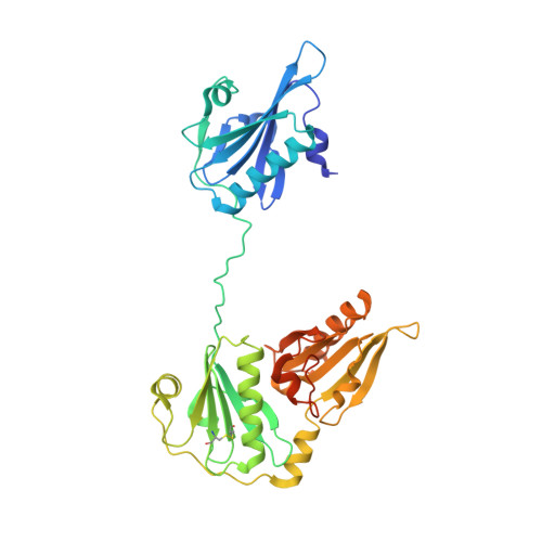
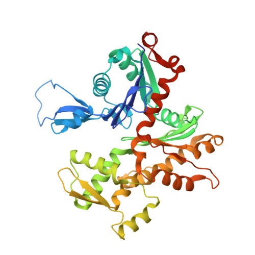
Ca2+ binding by domain 2 plays a critical role in the activation and stabilization of gelsolin.

Nag, S.Ma, Q.Wang, H.Chumnarnsilpa, S.Lee, W.L.Larsson, M.Kannan, B.Hernandez-Valladares, M.Burtnick, L.D.Robinson, R.C.

(2009) Proc Natl Acad Sci U S A 106: 13713-13718

  • DOI: https://doi.org/10.1073/pnas.0812374106
  • Primary Citation of Related Structures:  
    3FFK, 3FFN

  • PubMed Abstract: 
  • Gelsolin consists of six homologous domains (G1-G6), each containing a conserved Ca-binding site. Occupation of a subset of these sites enables gelsolin to sever and cap actin filaments in a Ca-dependent manner. Here, we present the structures of Ca-free human gelsolin and of Ca-bound human G1-G3 in a complex with actin ...

    Gelsolin consists of six homologous domains (G1-G6), each containing a conserved Ca-binding site. Occupation of a subset of these sites enables gelsolin to sever and cap actin filaments in a Ca-dependent manner. Here, we present the structures of Ca-free human gelsolin and of Ca-bound human G1-G3 in a complex with actin. These structures closely resemble those determined previously for equine gelsolin. However, the G2 Ca-binding site is occupied in the human G1-G3/actin structure, whereas it is vacant in the equine version. In-depth comparison of the Ca-free and Ca-activated, actin-bound human gelsolin structures suggests G2 and G6 to be cooperative in binding Ca(2+) and responsible for opening the G2-G6 latch to expose the F-actin-binding site on G2. Mutational analysis of the G2 and G6 Ca-binding sites demonstrates their interdependence in maintaining the compact structure in the absence of calcium. Examination of Ca binding by G2 in human G1-G3/actin reveals that the Ca(2+) locks the G2-G3 interface. Thermal denaturation studies of G2-G3 indicate that Ca binding stabilizes this fragment, driving it into the active conformation. The G2 Ca-binding site is mutated in gelsolin from familial amyloidosis (Finnish-type) patients. This disease initially proceeds through protease cleavage of G2, ultimately to produce a fragment that forms amyloid fibrils. The data presented here support a mechanism whereby the loss of Ca binding by G2 prolongs the lifetime of partially activated, intermediate conformations in which the protease cleavage site is exposed.


    Organizational Affiliation

    Institute of Molecular and Cell Biology, ASTAR, 61 Biopolis Drive, Proteos, Singapore 138673.


Macromolecules
Find similar proteins by:  (by identity cutoff)  |  3D Structure
Entity ID: 1
MoleculeChainsSequence LengthOrganismDetailsImage
plasma gelsolinA,
C [auth D]
377Homo sapiensMutation(s): 0 
Gene Names: GSN
UniProt & NIH Common Fund Data Resources
Find proteins for P06396 (Homo sapiens)
Explore P06396 
Go to UniProtKB:  P06396
PHAROS:  P06396
Entity Groups  
Sequence Clusters30% Identity50% Identity70% Identity90% Identity95% Identity100% Identity
UniProt GroupP06396
Protein Feature View
Expand
  • Reference Sequence
Find similar proteins by:  (by identity cutoff)  |  3D Structure
Entity ID: 2
MoleculeChainsSequence LengthOrganismDetailsImage
actin, alpha skeletal muscleB,
D [auth E]
377Oryctolagus cuniculusMutation(s): 0 
Gene Names: ACTA1ACTA
UniProt
Find proteins for P68135 (Oryctolagus cuniculus)
Explore P68135 
Go to UniProtKB:  P68135
Entity Groups  
Sequence Clusters30% Identity50% Identity70% Identity90% Identity95% Identity100% Identity
UniProt GroupP68135
Protein Feature View
Expand
  • Reference Sequence
Experimental Data & Validation

Experimental Data

  • Method: X-RAY DIFFRACTION
  • Resolution: 3.00 Å
  • R-Value Free: 0.273 
  • R-Value Work: 0.201 
  • R-Value Observed: 0.204 
  • Space Group: P 21 21 21
Unit Cell:
Length ( Å )Angle ( ˚ )
a = 102.179α = 90
b = 146.923β = 90
c = 148.301γ = 90
Software Package:
Software NamePurpose
DENZOdata reduction
SCALEPACKdata scaling
AMoREphasing
REFMACrefinement
PDB_EXTRACTdata extraction

Structure Validation

View Full Validation Report


Ligand Structure Quality Assessment 


Entry History 

Deposition Data

Revision History  (Full details and data files)

  • Version 1.0: 2009-10-06
    Type: Initial release
  • Version 1.1: 2011-07-13
    Changes: Advisory, Refinement description, Version format compliance
  • Version 1.2: 2017-11-01
    Changes: Refinement description