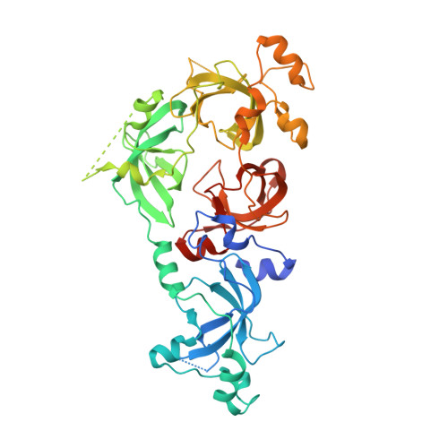
Structural studies of a four-MBT repeat protein MBTD1.
Eryilmaz, J., Pan, P., Amaya, M.F., Allali-Hassani, A., Dong, A., Adams-Cioaba, M.A., Mackenzie, F., Vedadi, M., Min, J.(2009) PLoS One 4: e7274-e7274
- PubMed: 19841675 Search on PubMedSearch on PubMed Central
- DOI: https://doi.org/10.1371/journal.pone.0007274
- Primary Citation Related Structures:
3FEO - PubMed Abstract:
The Polycomb group (PcG) of proteins is a family of important developmental regulators. The respective members function as large protein complexes involved in establishment and maintenance of transcriptional repression of developmental control genes. MBTD1, Malignant Brain Tumor domain-containing protein 1, is one such PcG protein. MBTD1 contains four MBT repeats. We have determined the crystal structure of MBTD1 (residues 130-566aa covering the 4 MBT repeats) at 2.5 A resolution by X-ray crystallography. The crystal structure of MBTD1 reveals its similarity to another four-MBT-repeat protein L3MBTL2, which binds lower methylated lysine histones. Fluorescence polarization experiments confirmed that MBTD1 preferentially binds mono- and di-methyllysine histone peptides, like L3MBTL1 and L3MBTL2. All known MBT-peptide complex structures characterized to date do not exhibit strong histone peptide sequence selectivity, and use a "cavity insertion recognition mode" to recognize the methylated lysine with the deeply buried methyl-lysine forming extensive interactions with the protein while the peptide residues flanking methyl-lysine forming very few contacts [1]. Nevertheless, our mutagenesis data based on L3MBTL1 suggested that the histone peptides could not bind to MBT repeats in any orientation. The four MBT repeats in MBTD1 exhibits an asymmetric rhomboid architecture. Like other MBT repeat proteins characterized so far, MBTD1 binds mono- or dimethylated lysine histones through one of its four MBT repeats utilizing a semi-aromatic cage. This article can also be viewed as an enhanced version in which the text of the article is integrated with interactive 3D representations and animated transitions. Please note that a web plugin is required to access this enhanced functionality. Instructions for the installation and use of the web plugin are available in Text S1.
- Structural Genomics Consortium, University of Toronto, Toronto, Ontario, Canada.
Organizational Affiliation: