3FE5

Crystal structure of 3-hydroxyanthranilate 3,4-dioxygenase from bovine kidney


Experimental Data Snapshot

  • Method: X-RAY DIFFRACTION
  • Resolution: 2.51 Å
  • R-Value Free: 0.240 
  • R-Value Work: 0.163 
  • R-Value Observed: 0.167 

wwPDB Validation   3D Report Full Report


This is version 1.4 of the entry. See complete history


Literature

Crystal structure of bovine 3-hydroxyanthranilate 3,4-dioxygenase.

Dilovic, I.Gliubich, F.Malpeli, G.Zanotti, G.Matkovic-Calogovic, D.

(2009) Biopolymers 

  • DOI: https://doi.org/10.1002/bip.21167
  • Primary Citation of Related Structures:  
    3FE5

  • PubMed Abstract: 

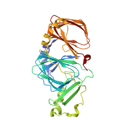
    3-Hydroxyanthranilate 3,4-dioxygenase, the enzyme that catalyzes the conversion of 3-hydroxyanthranilate to quinolinic acid, has been extracted and purified from bovine kidney, crystallized and its structure determined at 2.5 A resolution. The enzyme, which crystallizes in the triclinic P1 space group, is a monomer, characterized by the so-called cupin fold. The monomer of the bovine enzyme mimics the dimer present in lower species, such as bacteria and yeast, since it is composed of two domains: one of them is equivalent to one monomer, whilst the second domain corresponds to only a portion of it. The active site consists of an iron ion coordinated by two histidine residues, one glutamate and an external ligand, which has been interpreted as a solvent molecule. It is contained in the N-terminal domain, whilst the function of the C-terminal domain is possibly structural. The catalytic mechanism very likely has been conserved through all species, since the positions of all residues considered relevant for the reaction are present from bacteria to humans.


  • Organizational Affiliation

    Department of Chemistry, University of Zagreb, Horvatovac 102a, Zagreb 10000, Croatia.


Macromolecules
Find similar proteins by:  (by identity cutoff)  |  3D Structure
Entity ID: 1
MoleculeChains Sequence LengthOrganismDetailsImage
3-hydroxyanthranilate 3,4-dioxygenase285Bos taurusMutation(s): 0 
EC: 1.13.11.6
UniProt
Find proteins for Q0VCA8 (Bos taurus)
Explore Q0VCA8 
Go to UniProtKB:  Q0VCA8
Entity Groups  
Sequence Clusters30% Identity50% Identity70% Identity90% Identity95% Identity100% Identity
UniProt GroupQ0VCA8
Sequence Annotations
Expand
  • Reference Sequence
Small Molecules
Ligands 1 Unique
IDChains Name / Formula / InChI Key2D Diagram3D Interactions
FE
Query on FE

Download Ideal Coordinates CCD File 
B [auth A]FE (III) ION
Fe
VTLYFUHAOXGGBS-UHFFFAOYSA-N
Modified Residues  1 Unique
IDChains TypeFormula2D DiagramParent
CSO
Query on CSO
A
L-PEPTIDE LINKINGC3 H7 N O3 SCYS
Experimental Data & Validation

Experimental Data

  • Method: X-RAY DIFFRACTION
  • Resolution: 2.51 Å
  • R-Value Free: 0.240 
  • R-Value Work: 0.163 
  • R-Value Observed: 0.167 
  • Space Group: P 1
Unit Cell:
Length ( Å )Angle ( ˚ )
a = 40.25α = 91.7
b = 42.52β = 113.3
c = 46.48γ = 107.7
Software Package:
Software NamePurpose
MAR345data collection
AMoREphasing
REFMACrefinement
XDSdata reduction

Structure Validation

View Full Validation Report


Entry History 

Deposition Data

Revision History  (Full details and data files)

  • Version 1.0: 2009-06-09
    Type: Initial release
  • Version 1.1: 2011-07-13
    Changes: Version format compliance
  • Version 1.2: 2017-11-01
    Changes: Refinement description
  • Version 1.3: 2023-11-01
    Changes: Data collection, Database references, Derived calculations, Refinement description
  • Version 1.4: 2023-11-15
    Changes: Data collection