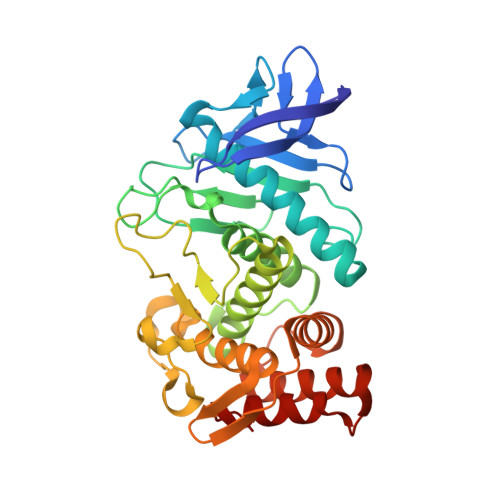
3FCQ

Thermolysin inhibition


Experimental Data Snapshot

  • Method: X-RAY DIFFRACTION
  • Resolution: 1.75 Å
  • R-Value Free: 0.231 
  • R-Value Work: 0.176 
  • R-Value Observed: 0.177 

Starting Model: experimental
View more details

wwPDB Validation   3D Report Full Report


Ligand Structure Quality Assessment 


This is version 1.3 of the entry. See complete history


Literature

Fragment-Based Lead Discovery: Screening and Optimizing Fragments for Thermolysin Inhibition.

Englert, L.Silber, K.Steuber, H.Brass, S.Over, B.Gerber, H.D.Heine, A.Diederich, W.E.Klebe, G.

(2010) ChemMedChem 5: 930-940

  • DOI: https://doi.org/10.1002/cmdc.201000084
  • Primary Citation of Related Structures:  
    3F28, 3F2P, 3FCQ

  • PubMed Abstract: 

    Fragment-based drug discovery has gained a foothold in today's lead identification processes. We present the application of in silico fragment-based screening for the discovery of novel lead compounds for the metalloendoproteinase thermolysin. We have chosen thermolysin to validate our screening approach as it is a well-studied enzyme and serves as a model system for other proteases. A protein-targeted virtual library was designed and screening was carried out using the program AutoDock. Two fragment hits could be identified. For one of them, the crystal structure in complex with thermolysin is presented. This compound was selected for structure-based optimization of binding affinity and improvement of ligand efficiency, while concomitantly keeping the fragment-like properties of the initial hit. Redesigning the zinc coordination group revealed a novel class of fragments possessing K(i) values as low as 128 microM, thus they provide a good starting point for further hit evolution in a tailored lead design.


  • Organizational Affiliation

    Philipps-Universität Marburg, Institut für Pharmazeutische Chemie, Marbacher Weg 6, 35032 Marburg, Germany.


Macromolecules
Find similar proteins by:  (by identity cutoff)  |  3D Structure
Entity ID: 1
MoleculeChains Sequence LengthOrganismDetailsImage
Thermolysin316Bacillus thermoproteolyticusMutation(s): 2 
EC: 3.4.24.27
UniProt
Find proteins for P00800 (Bacillus thermoproteolyticus)
Explore P00800 
Go to UniProtKB:  P00800
Entity Groups  
Sequence Clusters30% Identity50% Identity70% Identity90% Identity95% Identity100% Identity
UniProt GroupP00800
Sequence Annotations
Expand
  • Reference Sequence
Small Molecules
Binding Affinity Annotations 
IDSourceBinding Affinity
M3S PDBBind:  3FCQ Ki: 1.70e+6 (nM) from 1 assay(s)
Experimental Data & Validation

Experimental Data

  • Method: X-RAY DIFFRACTION
  • Resolution: 1.75 Å
  • R-Value Free: 0.231 
  • R-Value Work: 0.176 
  • R-Value Observed: 0.177 
  • Space Group: P 61 2 2
Unit Cell:
Length ( Å )Angle ( ˚ )
a = 92.718α = 90
b = 92.718β = 90
c = 129.965γ = 120
Software Package:
Software NamePurpose
SHELXL-97refinement
CNSrefinement
MAR345dtbdata collection
HKL-2000data reduction
HKL-2000data scaling
CNSphasing

Structure Validation

View Full Validation Report


Ligand Structure Quality Assessment 


Entry History 

Deposition Data

Revision History  (Full details and data files)

  • Version 1.0: 2009-12-08
    Type: Initial release
  • Version 1.1: 2011-07-13
    Changes: Version format compliance
  • Version 1.2: 2019-07-24
    Changes: Data collection, Refinement description
  • Version 1.3: 2023-09-06
    Changes: Data collection, Database references, Derived calculations, Refinement description