3F6K

Crystal structure of the Vps10p domain of human sortilin/NTS3 in complex with neurotensin


Experimental Data Snapshot

  • Method: X-RAY DIFFRACTION
  • Resolution: 2.00 Å
  • R-Value Free: 0.229 
  • R-Value Work: 0.204 
  • R-Value Observed: 0.205 

wwPDB Validation   3D Report Full Report


This is version 3.0 of the entry. See complete history


Literature

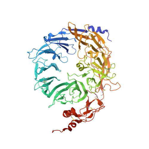
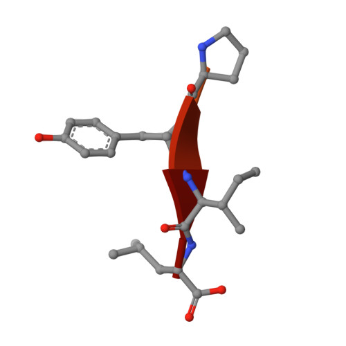
Ligands bind to Sortilin in the tunnel of a ten-bladed beta-propeller domain.

Quistgaard, E.M.Madsen, P.Groftehauge, M.K.Nissen, P.Petersen, C.M.Thirup, S.S.

(2009) Nat Struct Mol Biol 16: 96-98

  • DOI: https://doi.org/10.1038/nsmb.1543
  • Primary Citation of Related Structures:  
    3F6K

  • PubMed Abstract: 
  • The structure of the Sortilin ectodomain in complex with neurotensin has been determined at 2-A resolution, revealing that the C-terminal part of neurotensin binds in the tunnel of a ten-bladed beta-propeller domain. Binding competition studies suggest that additional binding sites, for example, for the prodomain of nerve growth factor-beta, are present in the tunnel and that competition for binding relates to the restricted space inside the propeller ...

    The structure of the Sortilin ectodomain in complex with neurotensin has been determined at 2-A resolution, revealing that the C-terminal part of neurotensin binds in the tunnel of a ten-bladed beta-propeller domain. Binding competition studies suggest that additional binding sites, for example, for the prodomain of nerve growth factor-beta, are present in the tunnel and that competition for binding relates to the restricted space inside the propeller.


    Organizational Affiliation

    MIND Centre, Department of Molecular Biology, University of Aarhus, Gustav Wieds Vej 10C, DK 8000 Arhus C, Denmark.


Macromolecules
Find similar proteins by:  (by identity cutoff)  |  3D Structure
Entity ID: 1
MoleculeChainsSequence LengthOrganismDetailsImage
Sortilin685Homo sapiensMutation(s): 1 
Gene Names: SORT1
UniProt & NIH Common Fund Data Resources
Find proteins for Q99523 (Homo sapiens)
Explore Q99523 
Go to UniProtKB:  Q99523
PHAROS:  Q99523
Entity Groups  
Sequence Clusters30% Identity50% Identity70% Identity90% Identity95% Identity100% Identity
UniProt GroupQ99523
Protein Feature View
Expand
  • Reference Sequence

Find similar proteins by:  Sequence   |   3D Structure  

Entity ID: 2
MoleculeChainsSequence LengthOrganismDetailsImage
NeurotensinB [auth N]13N/AMutation(s): 0 
UniProt & NIH Common Fund Data Resources
Find proteins for P30990 (Homo sapiens)
Explore P30990 
Go to UniProtKB:  P30990
PHAROS:  P30990
Entity Groups  
Sequence Clusters30% Identity50% Identity70% Identity90% Identity95% Identity100% Identity
UniProt GroupP30990
Protein Feature View
Expand
  • Reference Sequence
Oligosaccharides

Help

Entity ID: 3
MoleculeChainsChain Length2D DiagramGlycosylation3D Interactions
2-acetamido-2-deoxy-beta-D-glucopyranose-(1-4)-2-acetamido-2-deoxy-beta-D-glucopyranoseC [auth B]2N-Glycosylation Oligosaccharides Interaction
Glycosylation Resources
GlyTouCan:  G42666HT
GlyCosmos:  G42666HT
GlyGen:  G42666HT
Entity ID: 4
MoleculeChainsChain Length2D DiagramGlycosylation3D Interactions
beta-D-mannopyranose-(1-4)-2-acetamido-2-deoxy-beta-D-glucopyranose-(1-4)-2-acetamido-2-deoxy-beta-D-glucopyranoseD [auth C]3N-Glycosylation Oligosaccharides Interaction
Glycosylation Resources
GlyTouCan:  G15407YE
GlyCosmos:  G15407YE
GlyGen:  G15407YE
Small Molecules
Ligands 2 Unique
IDChainsName / Formula / InChI Key2D Diagram3D Interactions
PGE
Query on PGE

Download Ideal Coordinates CCD File 
E [auth A]TRIETHYLENE GLYCOL
C6 H14 O4
ZIBGPFATKBEMQZ-UHFFFAOYSA-N
 Ligand Interaction
GOL
Query on GOL

Download Ideal Coordinates CCD File 
F [auth A]GLYCEROL
C3 H8 O3
PEDCQBHIVMGVHV-UHFFFAOYSA-N
 Ligand Interaction
Modified Residues  1 Unique
IDChainsTypeFormula2D DiagramParent
PCA
Query on PCA
B [auth N]L-PEPTIDE LINKINGC5 H7 N O3GLN
Experimental Data & Validation

Experimental Data

  • Method: X-RAY DIFFRACTION
  • Resolution: 2.00 Å
  • R-Value Free: 0.229 
  • R-Value Work: 0.204 
  • R-Value Observed: 0.205 
  • Space Group: C 1 2 1
Unit Cell:
Length ( Å )Angle ( ˚ )
a = 145.76α = 90
b = 74.53β = 131.87
c = 108.33γ = 90
Software Package:
Software NamePurpose
XDSdata scaling
SHARPphasing
REFMACrefinement
XDSdata reduction
XSCALEdata scaling

Structure Validation

View Full Validation Report


Entry History 

Deposition Data

Revision History  (Full details and data files)

  • Version 1.0: 2008-12-30
    Type: Initial release
  • Version 1.1: 2011-07-13
    Changes: Advisory, Version format compliance
  • Version 2.0: 2019-12-25
    Changes: Database references, Derived calculations, Polymer sequence
  • Version 3.0: 2020-07-29
    Type: Remediation
    Reason: Carbohydrate remediation
    Changes: Atomic model, Data collection, Derived calculations, Structure summary