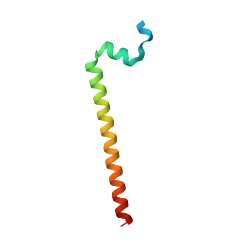
3F5H

Crystal structure of fused docking domains from PikAIII and PikAIV of the pikromycin polyketide synthase


Experimental Data Snapshot

  • Method: X-RAY DIFFRACTION
  • Resolution: 1.75 Å
  • R-Value Free: 0.250 
  • R-Value Work: 0.201 
  • R-Value Observed: 0.203 

wwPDB Validation   3D Report Full Report


This is version 1.4 of the entry. See complete history


Literature

Structural basis for binding specificity between subclasses of modular polyketide synthase docking domains.

Buchholz, T.J.Geders, T.W.Bartley, F.E.Reynolds, K.A.Smith, J.L.Sherman, D.H.

(2009) ACS Chem Biol 4: 41-52

  • DOI: https://doi.org/10.1021/cb8002607
  • Primary Citation of Related Structures:  
    3F5H

  • PubMed Abstract: 

    Bacterial type I polyketide synthases (PKSs) assemble structurally diverse natural products of significant clinical value from simple metabolic building blocks. The synthesis of these compounds occurs in a processive fashion along a large multiprotein complex. Transfer of the acyl intermediate across interpolypeptide junctions is mediated, at least in large part, by N- and C-terminal docking domains. We report here a comprehensive analysis of the binding affinity and selectivity for the complete set of discrete docking domain pairs in the pikromycin and erythromycin PKS systems. Despite disconnection from their parent module, each cognate pair of docking domains retained exquisite binding selectivity. Further insights were obtained by X-ray crystallographic analysis of the PikAIII/PikAIV docking domain interface. This new information revealed a series of key interacting residues that enabled development of a structural model for the recently proposed H2-T2 class of polypeptides involved in PKS intermodular molecular recognition.


  • Organizational Affiliation

    Life Sciences Institute, Department of Medicinal Chemistry, University of Michigan, Ann Arbor, Michigan 48109, USA.


Macromolecules
Find similar proteins by:  (by identity cutoff)  |  3D Structure
Entity ID: 1
MoleculeChains Sequence LengthOrganismDetailsImage
Type I polyketide synthase PikAIII, Type I polyketide synthase PikAIV fusion protein
A, B
68Streptomyces venezuelaeMutation(s): 0 
Gene Names: pIKAIIIpIKAIV
UniProt
Find proteins for Q9ZGI3 (Streptomyces venezuelae)
Explore Q9ZGI3 
Go to UniProtKB:  Q9ZGI3
Find proteins for Q9ZGI2 (Streptomyces venezuelae)
Explore Q9ZGI2 
Go to UniProtKB:  Q9ZGI2
Entity Groups  
Sequence Clusters30% Identity50% Identity70% Identity90% Identity95% Identity100% Identity
UniProt GroupsQ9ZGI3Q9ZGI2
Sequence Annotations
Expand
  • Reference Sequence
Small Molecules
Ligands 1 Unique
IDChains Name / Formula / InChI Key2D Diagram3D Interactions
NA
Query on NA

Download Ideal Coordinates CCD File 
C [auth B]SODIUM ION
Na
FKNQFGJONOIPTF-UHFFFAOYSA-N
Experimental Data & Validation

Experimental Data

  • Method: X-RAY DIFFRACTION
  • Resolution: 1.75 Å
  • R-Value Free: 0.250 
  • R-Value Work: 0.201 
  • R-Value Observed: 0.203 
  • Space Group: C 2 2 21
Unit Cell:
Length ( Å )Angle ( ˚ )
a = 59.016α = 90
b = 117.944β = 90
c = 41.773γ = 90
Software Package:
Software NamePurpose
DENZOdata reduction
SCALEPACKdata scaling
SOLVEphasing
RESOLVEphasing
REFMACrefinement
PDB_EXTRACTdata extraction
HKL-2000data collection

Structure Validation

View Full Validation Report


Entry History 

Deposition Data

Revision History  (Full details and data files)

  • Version 1.0: 2009-01-27
    Type: Initial release
  • Version 1.1: 2011-07-13
    Changes: Version format compliance
  • Version 1.2: 2017-08-02
    Changes: Refinement description, Source and taxonomy
  • Version 1.3: 2017-10-25
    Changes: Refinement description
  • Version 1.4: 2023-12-27
    Changes: Data collection, Database references, Derived calculations