3F5C | pdb_00003f5c

Structure of Dax-1:LRH-1 complex


Experimental Data Snapshot

  • Method: X-RAY DIFFRACTION
  • Resolution: 3.00 Å
  • R-Value Free: 
    0.279 (Depositor), 0.271 (DCC) 
  • R-Value Work: 
    0.222 (Depositor), 0.219 (DCC) 
  • R-Value Observed: 
    0.224 (Depositor) 

Starting Model: experimental
View more details

wwPDB Validation 3D Report Full Report

Validation slider image for 3F5C

This is version 1.4 of the entry. See complete history

Literature

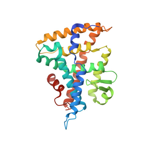
The structure of corepressor Dax-1 bound to its target nuclear receptor LRH-1.

Sablin, E.P.Woods, A.Krylova, I.N.Hwang, P.Ingraham, H.A.Fletterick, R.J.

(2008) Proc Natl Acad Sci U S A 105: 18390-18395

  • DOI: https://doi.org/10.1073/pnas.0808936105
  • Primary Citation Related Structures: 
    3F5C

  • PubMed Abstract: 

    The Dax-1 protein is an enigmatic nuclear receptor that lacks an expected DNA binding domain, yet functions as a potent corepressor of nuclear receptors. Here we report the structure of Dax-1 bound to one of its targets, liver receptor homolog 1 (LRH-1). Unexpectedly, Dax-1 binds to LRH-1 using a new module, a repressor helix built from a family conserved sequence motif, PCFXXLP. Mutations in this repressor helix that are linked with human endocrine disorders dissociate the complex and attenuate Dax-1 function. The structure of the Dax-1:LRH-1 complex provides the molecular mechanism for the function of Dax-1 as a potent transcriptional repressor.


  • Organizational Affiliation
    • Department of Biochemistry and Biophysics, University of California, San Francisco, CA 94143, USA.

Macromolecule Content 

  • Total Structure Weight: 89.48 kDa 
  • Atom Count: 4,936 
  • Modeled Residue Count: 607 
  • Deposited Residue Count: 784 
  • Unique protein chains: 2

Macromolecules

Find similar proteins by:|  3D Structure
Entity ID: 1
MoleculeChains  Sequence LengthOrganismDetailsImage
Nuclear receptor subfamily 5 group A member 2248Mus musculusMutation(s): 1 
Gene Names: Nr5a2Lrh1
UniProt
Find proteins for P45448 (Mus musculus)
Explore P45448 
Go to UniProtKB:  P45448
Entity Groups
Sequence Clusters30% Identity50% Identity70% Identity90% Identity95% Identity100% Identity
UniProt GroupP45448
Sequence Annotations
Expand
Reference Sequence
Find similar proteins by:|  3D Structure
Entity ID: 2
MoleculeChains  Sequence LengthOrganismDetailsImage
Nuclear receptor subfamily 0 group B member 1
B, C
268Mus musculusMutation(s): 0 
Gene Names: Nr0b1AhchDax1
UniProt & NIH Common Fund Data Resources
Find proteins for Q61066 (Mus musculus)
Explore Q61066 
Go to UniProtKB:  Q61066
IMPC:  MGI:1352460
Entity Groups
Sequence Clusters30% Identity50% Identity70% Identity90% Identity95% Identity100% Identity
UniProt GroupQ61066
Sequence Annotations
Expand
Reference Sequence

Experimental Data & Validation

Experimental Data

  • Method: X-RAY DIFFRACTION
  • Resolution: 3.00 Å
  • R-Value Free:  0.279 (Depositor), 0.271 (DCC) 
  • R-Value Work:  0.222 (Depositor), 0.219 (DCC) 
  • R-Value Observed: 0.224 (Depositor) 
Space Group: P 43
Unit Cell:
Length ( Å )Angle ( ˚ )
a = 103.355α = 90
b = 103.355β = 90
c = 117.455γ = 90
Software Package:
Software NamePurpose
PHENIXrefinement
CNSrefinement
ADSCdata collection
DENZOdata reduction
SCALEPACKdata scaling
CNSphasing

Structure Validation

View Full Validation Report


Entry History 

Deposition Data

Revision History  (Full details and data files)

  • Version 1.0: 2008-12-23
    Type: Initial release
  • Version 1.1: 2011-07-13
    Changes: Version format compliance
  • Version 1.2: 2019-07-24
    Changes: Data collection, Refinement description
  • Version 1.3: 2021-10-20
    Changes: Database references
  • Version 1.4: 2023-09-06
    Changes: Data collection, Refinement description