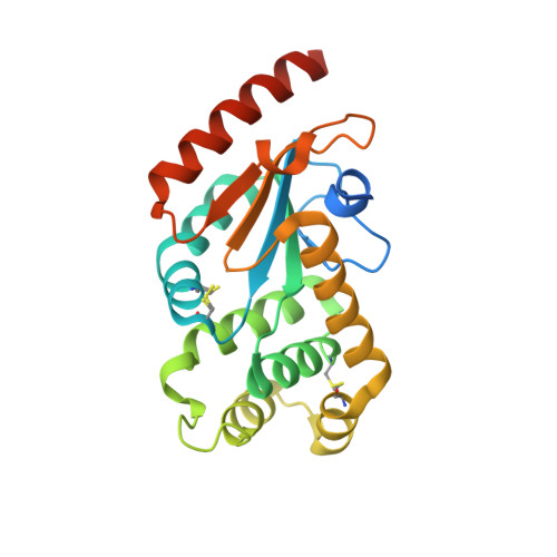
3F4R

Crystal structure of Wolbachia pipientis alpha-DsbA1


Experimental Data Snapshot

  • Method: X-RAY DIFFRACTION
  • Resolution: 1.60 Å
  • R-Value Free: 0.192 
  • R-Value Work: 0.165 
  • R-Value Observed: 0.168 

wwPDB Validation   3D Report Full Report


This is version 1.2 of the entry. See complete history


Literature

Structural and Functional Characterization of the Oxidoreductase alpha-DsbA1 from Wolbachia pipientis

Kurz, M.Iturbe-Ormaetxe, I.Jarrott, R.Shouldice, S.R.Wouters, M.A.Frei, P.Glockshuber, R.O'Neill, S.L.Heras, B.Martin, J.L.

(2009) Antioxid Redox Signal 11: 1485-1500

  • DOI: https://doi.org/10.1089/ars.2008.2420
  • Primary Citation of Related Structures:  
    3F4R, 3F4S, 3F4T

  • PubMed Abstract: 

    The alpha-proteobacterium Wolbachia pipientis is a highly successful intracellular endosymbiont of invertebrates that manipulates its host's reproductive biology to facilitate its own maternal transmission. The fastidious nature of Wolbachia and the lack of genetic transformation have hampered analysis of the molecular basis of these manipulations. Structure determination of key Wolbachia proteins will enable the development of inhibitors for chemical genetics studies. Wolbachia encodes a homologue (alpha-DsbA1) of the Escherichia coli dithiol oxidase enzyme EcDsbA, essential for the oxidative folding of many exported proteins. We found that the active-site cysteine pair of Wolbachia alpha-DsbA1 has the most reducing redox potential of any characterized DsbA. In addition, Wolbachia alpha-DsbA1 possesses a second disulfide that is highly conserved in alpha-proteobacterial DsbAs but not in other DsbAs. The alpha-DsbA1 structure lacks the characteristic hydrophobic features of EcDsbA, and the protein neither complements EcDsbA deletion mutants in E. coli nor interacts with EcDsbB, the redox partner of EcDsbA. The surface characteristics and redox profile of alpha-DsbA1 indicate that it probably plays a specialized oxidative folding role with a narrow substrate specificity. This first report of a Wolbachia protein structure provides the basis for future chemical genetics studies.


  • Organizational Affiliation

    Institute for Molecular Bioscience, University of New South Wales, Sydney, Australia.


Macromolecules
Find similar proteins by:  (by identity cutoff)  |  3D Structure
Entity ID: 1
MoleculeChains Sequence LengthOrganismDetailsImage
Putative uncharacterized protein226Wolbachia endosymbiont of Drosophila melanogasterMutation(s): 0 
Gene Names: WD_1055
UniProt
Find proteins for Q73GA4 (Wolbachia pipientis wMel)
Explore Q73GA4 
Go to UniProtKB:  Q73GA4
Entity Groups  
Sequence Clusters30% Identity50% Identity70% Identity90% Identity95% Identity100% Identity
UniProt GroupQ73GA4
Sequence Annotations
Expand
  • Reference Sequence
Small Molecules
Ligands 2 Unique
IDChains Name / Formula / InChI Key2D Diagram3D Interactions
1PE
Query on 1PE

Download Ideal Coordinates CCD File 
C [auth A]PENTAETHYLENE GLYCOL
C10 H22 O6
JLFNLZLINWHATN-UHFFFAOYSA-N
PGE
Query on PGE

Download Ideal Coordinates CCD File 
B [auth A]TRIETHYLENE GLYCOL
C6 H14 O4
ZIBGPFATKBEMQZ-UHFFFAOYSA-N
Experimental Data & Validation

Experimental Data

  • Method: X-RAY DIFFRACTION
  • Resolution: 1.60 Å
  • R-Value Free: 0.192 
  • R-Value Work: 0.165 
  • R-Value Observed: 0.168 
  • Space Group: C 1 2 1
Unit Cell:
Length ( Å )Angle ( ˚ )
a = 71.4α = 90
b = 49.6β = 107
c = 69.7γ = 90
Software Package:
Software NamePurpose
d*TREKdata scaling
PHENIXrefinement
PDB_EXTRACTdata extraction
d*TREKdata reduction
SCALEPACKdata scaling
SOLVEphasing

Structure Validation

View Full Validation Report


Entry History 

Deposition Data

Revision History  (Full details and data files)

  • Version 1.0: 2009-03-24
    Type: Initial release
  • Version 1.1: 2011-07-13
    Changes: Source and taxonomy, Version format compliance
  • Version 1.2: 2023-12-27
    Changes: Data collection, Database references, Derived calculations