3F3M | pdb_00003f3m

Six Crystal Structures of Two Phosphopantetheine Adenylyltransferases Reveal an Alternative Ligand Binding Mode and an Associated Structural Change


Experimental Data Snapshot

  • Method: X-RAY DIFFRACTION
  • Resolution: 2.40 Å
  • R-Value Free: 
    0.250 (Depositor), 0.277 (DCC) 
  • R-Value Work: 
    0.211 (Depositor) 

Starting Model: experimental
View more details

wwPDB Validation 3D Report Full Report

Validation slider image for 3F3M

Ligand Structure Quality Assessment 


This is version 1.3 of the entry. See complete history

Literature

The structure of Staphylococcus aureus phosphopantetheine adenylyltransferase in complex with 3'-phosphoadenosine 5'-phosphosulfate reveals a new ligand-binding mode

Lee, H.H.Yoon, H.J.Kang, J.Y.Park, J.H.Kim, D.J.Choi, K.H.Lee, S.K.Song, J.Kim, H.J.Suh, S.W.

(2009) Acta Crystallogr Sect F Struct Biol Cryst Commun 65: 987-991

  • DOI: https://doi.org/10.1107/S1744309109036616
  • Primary Citation Related Structures: 
    3F3M

  • PubMed Abstract: 

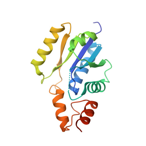
    Bacterial phosphopantetheine adenylyltransferase (PPAT) catalyzes the penultimate step in the coenzyme A (CoA) biosynthetic pathway. It catalyzes the reversible transfer of an adenylyl group from ATP to 4'-phosphopantetheine (Ppant) to form dephospho-CoA (dPCoA) and pyrophosphate. Previous structural studies have revealed how several ligands are recognized by bacterial PPATs. ATP, ADP, Ppant and dPCoA bind to the same binding site in a highly similar manner, while CoA binds to a partially overlapping site in a different mode. To provide further structural insights into ligand binding, the crystal structure of Staphylococcus aureus PPAT was solved in a binary complex with 3'-phosphoadenosine 5'-phosphosulfate (PAPS). This study unexpectedly revealed a new mode of ligand binding to PPAT, thus providing potentially useful information for structure-based discovery of inhibitors of bacterial PPATs.


  • Organizational Affiliation
    • Department of Chemistry, College of Natural Sciences, Seoul National University, Seoul 151-742, Republic of Korea.

Macromolecule Content 

  • Total Structure Weight: 19.98 kDa 
  • Atom Count: 1,323 
  • Modeled Residue Count: 155 
  • Deposited Residue Count: 168 
  • Unique protein chains: 1

Macromolecules

Find similar proteins by:|  3D Structure
Entity ID: 1
MoleculeChains  Sequence LengthOrganismDetailsImage
Phosphopantetheine adenylyltransferase168Staphylococcus aureusMutation(s): 0 
Gene Names: coaD
EC: 2.7.7.3
UniProt
Find proteins for P63820 (Staphylococcus aureus (strain MW2))
Explore P63820 
Go to UniProtKB:  P63820
Entity Groups
Sequence Clusters30% Identity50% Identity70% Identity90% Identity95% Identity100% Identity
UniProt GroupP63820
Sequence Annotations
Expand
Reference Sequence

Small Molecules

Ligands 1 Unique
IDChains Name / Formula / InChI Key2D Diagram3D Interactions
PPS

Query on PPS


Download:Ideal Coordinates CCD File
B [auth A]3'-PHOSPHATE-ADENOSINE-5'-PHOSPHATE SULFATE
C10 H15 N5 O13 P2 S
GACDQMDRPRGCTN-KQYNXXCUSA-N

Experimental Data & Validation

Experimental Data

  • Method: X-RAY DIFFRACTION
  • Resolution: 2.40 Å
  • R-Value Free:  0.250 (Depositor), 0.277 (DCC) 
  • R-Value Work:  0.211 (Depositor) 
Space Group: P 63 2 2
Unit Cell:
Length ( Å )Angle ( ˚ )
a = 66.774α = 90
b = 66.774β = 90
c = 124.134γ = 120
Software Package:
Software NamePurpose
CNSrefinement
PDB_EXTRACTdata extraction
HKL-2000data collection
HKL-2000data reduction
HKL-2000data scaling
CNSphasing

Structure Validation

View Full Validation Report


Ligand Structure Quality Assessment 


Entry History 

Deposition Data

Revision History  (Full details and data files)

  • Version 1.0: 2009-10-20
    Type: Initial release
  • Version 1.1: 2011-07-13
    Changes: Version format compliance
  • Version 1.2: 2012-04-25
    Changes: Database references
  • Version 1.3: 2023-11-01
    Changes: Data collection, Database references, Derived calculations, Refinement description