3F27 | pdb_00003f27

Structure of Sox17 Bound to DNA


Experimental Data Snapshot

  • Method: X-RAY DIFFRACTION
  • Resolution: 2.75 Å
  • R-Value Free: 
    0.265 (Depositor), 0.259 (DCC) 
  • R-Value Work: 
    0.242 (Depositor), 0.231 (DCC) 
  • R-Value Observed: 
    0.243 (Depositor) 

Starting Model: experimental
View more details

wwPDB Validation 3D Report Full Report

Validation slider image for 3F27

This is version 1.3 of the entry. See complete history

Literature

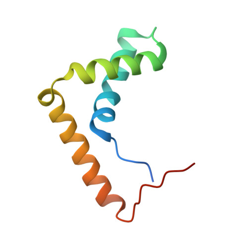
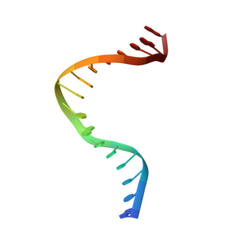
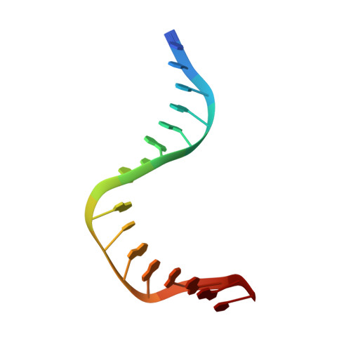
The Structure of Sox17 Bound to DNA Reveals a Conserved Bending Topology but Selective Protein Interaction Platforms

Palasingam, P.Jauch, R.Ng, C.K.L.Kolatkar, P.R.

(2009) J Mol Biology 388: 619-630

  • DOI: https://doi.org/10.1016/j.jmb.2009.03.055
  • Primary Citation Related Structures: 
    3F27

  • PubMed Abstract: 

    Sox17 regulates endodermal lineage commitment and is thought to function antagonistically to the pluripotency determinant Sox2. To investigate the biochemical basis for the distinct functions of Sox2 and Sox17, we solved the crystal structure of the high mobility group domain of Sox17 bound to a DNA element derived from the Lama1 enhancer using crystals diffracting to 2.7 A resolution. Sox17 targets the minor groove and bends the DNA by approximately 80 degrees . The DNA architecture closely resembles the one seen for Sox2/DNA structures, suggesting that the degree of bending is conserved between both proteins and nucleotide substitutions have only marginal effects on the bending topology. Accordingly, affinities of Sox2 and Sox17 for the Lama1 element were found to be identical. However, when the Oct1 contact interface of Sox2 is compared with the corresponding region of Sox17, a significantly altered charge distribution is observed, suggesting differential co-factor recruitment that may explain their biological distinctiveness.


  • Organizational Affiliation
    • Laboratory of Structural Biochemistry, Genome Institute of Singapore, 60 Biopolis Street, Singapore, Singapore.

Macromolecule Content 

  • Total Structure Weight: 19.88 kDa 
  • Atom Count: 1,283 
  • Modeled Residue Count: 106 
  • Deposited Residue Count: 115 
  • Unique protein chains: 1
  • Unique nucleic acid chains: 2

Macromolecules


Find similar proteins by:|  3D Structure
Entity ID: 3
MoleculeChains  Sequence LengthOrganismDetailsImage
Transcription factor SOX-17C [auth D]83Mus musculusMutation(s): 0 
Gene Names: Sox-17Sox17
UniProt
Find proteins for Q61473 (Mus musculus)
Explore Q61473 
Go to UniProtKB:  Q61473
Entity Groups
Sequence Clusters30% Identity50% Identity70% Identity90% Identity95% Identity100% Identity
UniProt GroupQ61473
Sequence Annotations
Expand
Reference Sequence
Find similar nucleic acids by:  Sequence
Entity ID: 1
MoleculeChains LengthOrganismImage
DNA (5'-D(*DGP*DTP*DCP*DTP*DCP*DTP*DAP*DTP*DTP*DGP*DTP*DCP*DCP*DTP*DGP*DG)-3')16synthetic construct
Sequence Annotations
Expand
Reference Sequence
Find similar nucleic acids by:  Sequence
Entity ID: 2
MoleculeChains LengthOrganismImage
DNA (5'-D(*DCP*DCP*DAP*DGP*DGP*DAP*DCP*DAP*DAP*DTP*DAP*DGP*DAP*DGP*DAP*DC)-3')16synthetic construct
Sequence Annotations
Expand
Reference Sequence

Experimental Data & Validation

Experimental Data

  • Method: X-RAY DIFFRACTION
  • Resolution: 2.75 Å
  • R-Value Free:  0.265 (Depositor), 0.259 (DCC) 
  • R-Value Work:  0.242 (Depositor), 0.231 (DCC) 
  • R-Value Observed: 0.243 (Depositor) 
Space Group: P 32 2 1
Unit Cell:
Length ( Å )Angle ( ˚ )
a = 68.753α = 90
b = 68.753β = 90
c = 66.329γ = 120
Software Package:
Software NamePurpose
SAINTdata scaling
REFMACrefinement
PDB_EXTRACTdata extraction

Structure Validation

View Full Validation Report


Entry History 

Deposition Data

Revision History  (Full details and data files)

  • Version 1.0: 2009-04-07
    Type: Initial release
  • Version 1.1: 2011-07-13
    Changes: Advisory, Version format compliance
  • Version 1.2: 2023-06-14
    Changes: Database references, Derived calculations, Source and taxonomy
  • Version 1.3: 2023-11-08
    Changes: Data collection, Refinement description