3EXS

Crystal structure of KGPDC from Streptococcus mutans in complex with D-R5P


Experimental Data Snapshot

  • Method: X-RAY DIFFRACTION
  • Resolution: 2.50 Å
  • R-Value Free: 0.244 
  • R-Value Work: 0.184 
  • R-Value Observed: 0.187 

wwPDB Validation   3D Report Full Report


Ligand Structure Quality Assessment 


This is version 1.1 of the entry. See complete history


Literature

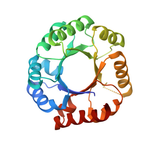
Open-closed conformational change revealed by the crystal structures of 3-keto-L-gulonate 6-phosphate decarboxylase from Streptococcus mutans

Li, G.L.Liu, X.Nan, J.Brostromer, E.Li, L.F.Su, X.D.

(2009) Biochem Biophys Res Commun 381: 429-433

  • DOI: https://doi.org/10.1016/j.bbrc.2009.02.049
  • Primary Citation of Related Structures:  
    3EXR, 3EXS, 3EXT

  • PubMed Abstract: 
  • The 3-keto-L-gulonate 6-phosphate decarboxylase (KGPDC) catalyses the decarboxylation of 3-keto-L-gulonate 6-phosphate to L-xylulose in the presence of magnesium ions. The enzyme is involved in L-ascorbate metabolism and plays an essential role in the pathway of glucuronate interconversion ...

    The 3-keto-L-gulonate 6-phosphate decarboxylase (KGPDC) catalyses the decarboxylation of 3-keto-L-gulonate 6-phosphate to L-xylulose in the presence of magnesium ions. The enzyme is involved in L-ascorbate metabolism and plays an essential role in the pathway of glucuronate interconversion. Crystal structures of Streptococcus mutans KGPDC were determined in the absence and presence of the product analog D-ribulose 5-phosphate. We have observed an 8 A alphaB-helix movement and other structural rearrangements around the active site between the apo-structures and product analog bound structure. These drastic conformational changes upon ligand binding are the first observation of this kind for the KGPDC family. The flexibilities of both the alpha-helix lid and the side chains of Arg144 and Arg197 are associated with substrate binding and product releasing. The open-closed conformational changes of the active site, through the movements of the alpha-helix lid and the arginine residues are important for substrate binding and catalysis.


    Organizational Affiliation

    The National Laboratory of Protein Engineering and Plant Genetic Engineering, College of Life Science, Peking University, Beijing 100871, People's Republic of China.


Macromolecules
Find similar proteins by:  (by identity cutoff)  |  3D Structure
Entity ID: 1
MoleculeChainsSequence LengthOrganismDetailsImage
RmpD (Hexulose-6-phosphate synthase)
A, B, C, D
221Streptococcus mutansMutation(s): 0 
Gene Names: ulaDrmpD
EC: 4.1.1.85
UniProt
Find proteins for Q93DA8 (Streptococcus mutans)
Explore Q93DA8 
Go to UniProtKB:  Q93DA8
Entity Groups  
Sequence Clusters30% Identity50% Identity70% Identity90% Identity95% Identity100% Identity
UniProt GroupQ93DA8
Protein Feature View
Expand
  • Reference Sequence
Small Molecules
Ligands 1 Unique
IDChainsName / Formula / InChI Key2D Diagram3D Interactions
5RP
Query on 5RP

Download Ideal Coordinates CCD File 
E [auth A],
F [auth D]
RIBULOSE-5-PHOSPHATE
C5 H11 O8 P
FNZLKVNUWIIPSJ-UHNVWZDZSA-N
 Ligand Interaction
Experimental Data & Validation

Experimental Data

  • Method: X-RAY DIFFRACTION
  • Resolution: 2.50 Å
  • R-Value Free: 0.244 
  • R-Value Work: 0.184 
  • R-Value Observed: 0.187 
  • Space Group: P 21 21 21
Unit Cell:
Length ( Å )Angle ( ˚ )
a = 59.152α = 90
b = 124.249β = 90
c = 130.066γ = 90
Software Package:
Software NamePurpose
SAINTdata scaling
REFMACrefinement
PDB_EXTRACTdata extraction

Structure Validation

View Full Validation Report


Ligand Structure Quality Assessment 


Entry History 

Deposition Data

Revision History  (Full details and data files)

  • Version 1.0: 2009-08-25
    Type: Initial release
  • Version 1.1: 2011-07-13
    Changes: Version format compliance