


Crystal Structure of D,D-heptose 1.7-bisphosphate phosphatase from E. Coli.
Taylor, P., Sugiman-Marangos, S.N., Zhang, K., DeLeon, G., Wright, G.D., Junop, M.S.To be published.
Experimental Data Snapshot
Starting Model: experimental
View more details
wwPDB Validation 3D Report Full Report

Entity ID: 1 | |||||
|---|---|---|---|---|---|
| Molecule | Chains | Sequence Length | Organism | Details | Image |
| D,D-heptose 1,7-bisphosphate phosphatase | 211 | Escherichia coli K-12 | Mutation(s): 0 Gene Names: b0200, gmhB, JW0196, yaeD EC: 3.1.3 (PDB Primary Data), 3.1.3.82 (UniProt) | 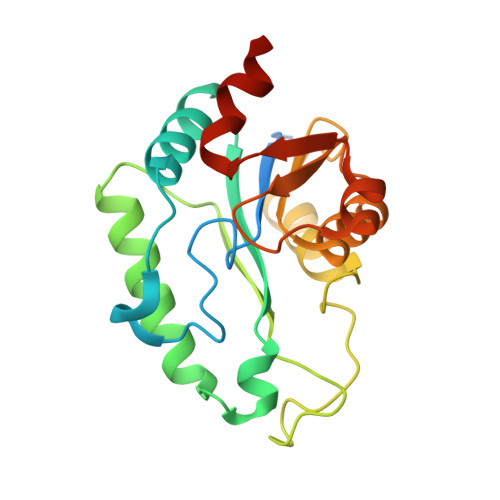 | |
UniProt | |||||
Entity Groups | |||||
| Sequence Clusters | 30% Identity50% Identity70% Identity90% Identity95% Identity100% Identity | ||||
| UniProt Group | P63228 | ||||
Sequence AnnotationsExpand | |||||
Reference Sequence | |||||
| Ligands 3 Unique | |||||
|---|---|---|---|---|---|
| ID | Chains | Name / Formula / InChI Key | 2D Diagram | 3D Interactions | |
| PO4 Download:Ideal Coordinates CCD File | D [auth A] | PHOSPHATE ION O4 P NBIIXXVUZAFLBC-UHFFFAOYSA-K |  | ||
| ZN Download:Ideal Coordinates CCD File | B [auth A] | ZINC ION Zn PTFCDOFLOPIGGS-UHFFFAOYSA-N |  | ||
| CA Download:Ideal Coordinates CCD File | C [auth A] | CALCIUM ION Ca BHPQYMZQTOCNFJ-UHFFFAOYSA-N |  | ||
| Length ( Å ) | Angle ( ˚ ) |
|---|---|
| a = 64.183 | α = 90 |
| b = 50.446 | β = 90 |
| c = 52.018 | γ = 90 |
| Software Name | Purpose |
|---|---|
| CrystalClear | data collection |
| PHENIX | model building |
| REFMAC | refinement |
| CrystalClear | data reduction |
| CrystalClear | data scaling |
| PHENIX | phasing |


