3ERS

Crystal Structure of E. coli Trbp111


Experimental Data Snapshot

  • Method: X-RAY DIFFRACTION
  • Resolution: 1.87 Å
  • R-Value Free: 0.282 
  • R-Value Work: 0.216 
  • R-Value Observed: 0.220 

wwPDB Validation   3D Report Full Report


This is version 1.3 of the entry. See complete history


Literature

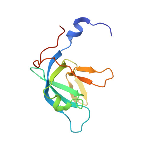
Crystal structure of Trbp111: a tructure specific tRNA binding protein

Swairjo, M.A.Morales, A.J.Wang, C.-C.Ortiz, A.R.Schimmel, P.

(2000) EMBO J 19: 6287-6298

  • DOI: https://doi.org/10.1093/emboj/19.23.6287
  • Primary Citation of Related Structures:  
    1PYB, 3ERS

  • PubMed Abstract: 

    Trbp111 is a 111 amino acid Aquifex aeolicus structure-specific tRNA-binding protein that has homologous counterparts distributed throughout evolution. A dimer is the functional unit for binding a single tRNA. Here we report the 3D structures of the A.aeolicus protein and its Escherichia coli homolog at resolutions of 2.50 and 1.87 A, respectively. The structure shows a symmetrical dimer of two core domains and a central dimerization domain where the N- and C-terminal regions of Trbp111 form an extensive dimer interface. The core of the monomer is a classical oligonucleotide/oligosaccharide-binding (OB) fold with a five-stranded ss-barrel and a small capping helix. This structure is similar to that seen in the anticodon-binding domain of three class II tRNA synthetases and several other proteins. Mutational analysis identified sites important for interactions with tRNA. These residues line the inner surfaces of two clefts formed between the ss-barrel of each monomer and the dimer interface. The results are consistent with a proposed model for asymmetrical docking of the convex side of tRNA to the dimer.


  • Organizational Affiliation

    Skaags Institute for Chemical Biology, Department of Molecular Biology and Department of Chemistry, The Scripps Research Institute, 10550 North Torrey Pines Road, La Jolla, CA 92037, USA.


Macromolecules
Find similar proteins by:  (by identity cutoff)  |  3D Structure
Entity ID: 1
MoleculeChains Sequence LengthOrganismDetailsImage
tRNA-binding protein ygjHA [auth X]118Escherichia coliMutation(s): 0 
Gene Names: ygjH
UniProt
Find proteins for P42589 (Escherichia coli (strain K12))
Explore P42589 
Go to UniProtKB:  P42589
Entity Groups  
Sequence Clusters30% Identity50% Identity70% Identity90% Identity95% Identity100% Identity
UniProt GroupP42589
Sequence Annotations
Expand
  • Reference Sequence
Experimental Data & Validation

Experimental Data

  • Method: X-RAY DIFFRACTION
  • Resolution: 1.87 Å
  • R-Value Free: 0.282 
  • R-Value Work: 0.216 
  • R-Value Observed: 0.220 
  • Space Group: P 32 2 1
Unit Cell:
Length ( Å )Angle ( ˚ )
a = 41.234α = 90
b = 41.234β = 90
c = 95.377γ = 120
Software Package:
Software NamePurpose
DENZOdata reduction
SCALEPACKdata scaling
SHARPphasing
SOLOMONphasing
REFMACrefinement
PDB_EXTRACTdata extraction
HKL-2000data reduction

Structure Validation

View Full Validation Report


Entry History 

Deposition Data

  • Released Date: 2008-12-02 
  • Deposition Author(s): Swairjo, M.A.
  • This entry supersedes: 1PXF

Revision History  (Full details and data files)

  • Version 1.0: 2008-12-02
    Type: Initial release
  • Version 1.1: 2011-07-13
    Changes: Version format compliance
  • Version 1.2: 2017-10-25
    Changes: Refinement description
  • Version 1.3: 2023-12-27
    Changes: Data collection, Database references