3ERE | pdb_00003ere

Crystal structure of the arginine repressor protein from Mycobacterium tuberculosis in complex with the DNA operator


Experimental Data Snapshot

  • Method: X-RAY DIFFRACTION
  • Resolution: 2.50 Å
  • R-Value Free: 
    0.255 (Depositor), 0.229 (DCC) 
  • R-Value Work: 
    0.227 (Depositor), 0.229 (DCC) 
  • R-Value Observed: 
    0.227 (Depositor) 

Starting Models: experimental
View more details

wwPDB Validation 3D Report Full Report

Validation slider image for 3ERE

This is version 1.2 of the entry. See complete history

Literature

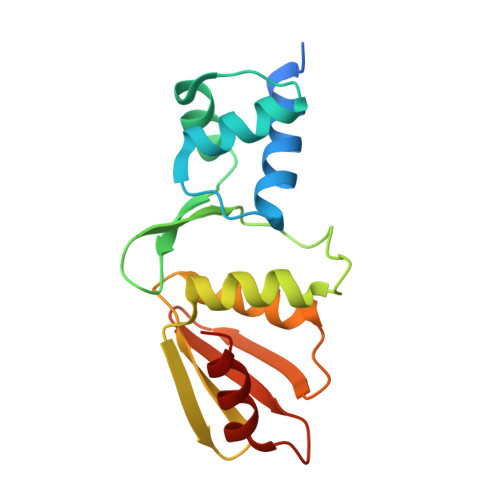
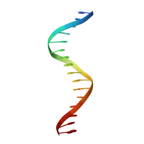
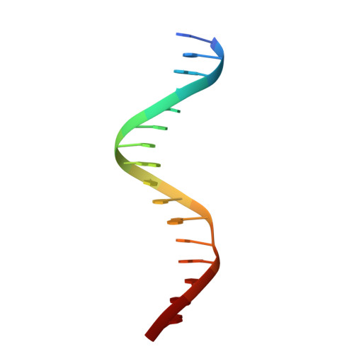
Crystal structure of the arginine repressor protein in complex with the DNA operator from Mycobacterium tuberculosis.

Cherney, L.T.Cherney, M.M.Garen, C.R.Lu, G.J.James, M.N.

(2008) J Mol Biology 384: 1330-1340

  • DOI: https://doi.org/10.1016/j.jmb.2008.10.015
  • Primary Citation Related Structures: 
    3ERE

  • PubMed Abstract: 

    The arginine repressor (ArgR) from Mycobacterium tuberculosis (Mtb) is a gene product encoded by the open reading frame Rv1657. It regulates the L-arginine concentration in cells by interacting with ARG boxes in the promoter regions of the arginine biosynthesis and catabolism operons. Here we present a 2.5-A structure of MtbArgR in complex with a 16-bp DNA operator in the absence of arginine. A biological trimer of the protein-DNA complex is formed via the crystallographic 3-fold symmetry axis. The N-terminal domain of MtbArgR has a winged helix-turn-helix motif that binds to the major groove of the DNA. This structure shows that, in the absence of arginine, the ArgR trimer can bind three ARG box half-sites. It also reveals the structure of the whole MtbArgR molecule itself containing both N-terminal and C-terminal domains.


  • Organizational Affiliation
    • Group in Protein Structure and Function, Department of Biochemistry, University of Alberta, 431 Medical Sciences Building, Edmonton, Alberta, Canada T6G 2H7.

Macromolecule Content 

  • Total Structure Weight: 27.45 kDa 
  • Atom Count: 1,880 
  • Modeled Residue Count: 188 
  • Deposited Residue Count: 202 
  • Unique protein chains: 1
  • Unique nucleic acid chains: 2

Macromolecules


Find similar proteins by:|  3D Structure
Entity ID: 1
MoleculeChains  Sequence LengthOrganismDetailsImage
Arginine repressorA [auth D]170Mycobacterium tuberculosis H37RvMutation(s): 0 
Gene Names: argRahrCRv1657MT1695MTCY06H11.22
UniProt
Find proteins for P9WPY9 (Mycobacterium tuberculosis (strain ATCC 25618 / H37Rv))
Explore P9WPY9 
Go to UniProtKB:  P9WPY9
Entity Groups
Sequence Clusters30% Identity50% Identity70% Identity90% Identity95% Identity100% Identity
UniProt GroupP9WPY9
Sequence Annotations
Expand
Reference Sequence
Find similar nucleic acids by:  Sequence
Entity ID: 2
MoleculeChains LengthOrganismImage
5'-D(*DTP*DTP*DGP*DCP*DAP*DTP*DAP*DAP*DCP*DGP*DAP*DTP*DGP*DCP*DAP*DA)-3'B [auth A]16N/A
Sequence Annotations
Expand
Reference Sequence
Find similar nucleic acids by:  Sequence
Entity ID: 3
MoleculeChains LengthOrganismImage
5'-D(*DTP*DTP*DGP*DCP*DAP*DTP*DCP*DGP*DTP*DTP*DAP*DTP*DGP*DCP*DAP*DA)-3'C [auth B]16N/A
Sequence Annotations
Expand
Reference Sequence

Experimental Data & Validation

Experimental Data

  • Method: X-RAY DIFFRACTION
  • Resolution: 2.50 Å
  • R-Value Free:  0.255 (Depositor), 0.229 (DCC) 
  • R-Value Work:  0.227 (Depositor), 0.229 (DCC) 
  • R-Value Observed: 0.227 (Depositor) 
Space Group: P 63 2 2
Unit Cell:
Length ( Å )Angle ( ˚ )
a = 94.128α = 90
b = 94.128β = 90
c = 147.014γ = 120
Software Package:
Software NamePurpose
ADSCdata collection
MOLREPphasing
CNSrefinement
HKL-2000data reduction
HKL-2000data scaling

Structure Validation

View Full Validation Report


Entry History 

Deposition Data

Revision History  (Full details and data files)

  • Version 1.0: 2008-10-14
    Type: Initial release
  • Version 1.1: 2011-07-13
    Changes: Source and taxonomy, Version format compliance
  • Version 1.2: 2023-09-06
    Changes: Data collection, Database references, Derived calculations, Refinement description