3EN1 | pdb_00003en1

Crystal structure of Toluene 2,3-Dioxygenase


Experimental Data Snapshot

  • Method: X-RAY DIFFRACTION
  • Resolution: 3.20 Å
  • R-Value Free: 
    0.208 (Depositor), 0.187 (DCC) 
  • R-Value Work: 
    0.185 (Depositor) 
  • R-Value Observed: 
    0.186 (Depositor) 

wwPDB Validation 3D Report Full Report

Validation slider image for 3EN1

This is version 1.4 of the entry. See complete history

Literature

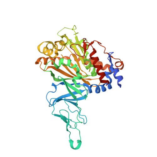
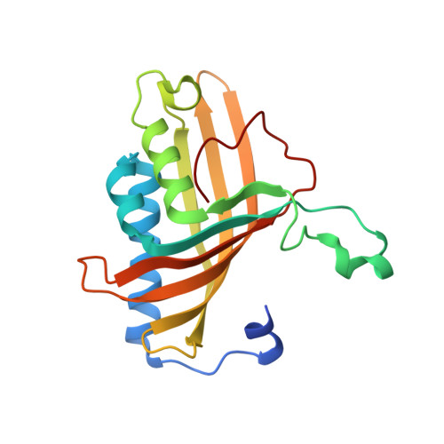
Structures of the multicomponent Rieske non-heme iron toluene 2,3-dioxygenase enzyme system

Friemann, R.Lee, K.Brown, E.N.Gibson, D.T.Eklund, H.Ramaswamy, S.

(2009) Acta Crystallogr D Biol Crystallogr 65: 24-33

  • DOI: https://doi.org/10.1107/S0907444908036524
  • Primary Citation Related Structures: 
    3DQY, 3EF6, 3EN1, 3EQQ

  • PubMed Abstract: 

    Bacterial Rieske non-heme iron oxygenases catalyze the initial hydroxylation of aromatic hydrocarbon substrates. The structures of all three components of one such system, the toluene 2,3-dioxygenase system, have now been determined. This system consists of a reductase, a ferredoxin and a terminal dioxygenase. The dioxygenase, which was cocrystallized with toluene, is a heterohexamer containing a catalytic and a structural subunit. The catalytic subunit contains a Rieske [2Fe-2S] cluster and mononuclear iron at the active site. This iron is not strongly bound and is easily removed during enzyme purification. The structures of the enzyme with and without mononuclear iron demonstrate that part of the structure is flexible in the absence of iron. The orientation of the toluene substrate in the active site is consistent with the regiospecificity of oxygen incorporation seen in the product formed. The ferredoxin is Rieske type and contains a [2Fe-2S] cluster close to the protein surface. The reductase belongs to the glutathione reductase family of flavoenzymes and consists of three domains: an FAD-binding domain, an NADH-binding domain and a C-terminal domain. A model for electron transfer from NADH via FAD in the reductase and the ferredoxin to the terminal active-site mononuclear iron of the dioxygenase is proposed.


  • Organizational Affiliation
    • Department of Molecular Biology, Swedish University of Agricultural Sciences, Box 590, 75124 Uppsala, Sweden.

Macromolecule Content 

  • Total Structure Weight: 74.06 kDa 
  • Atom Count: 4,917 
  • Modeled Residue Count: 604 
  • Deposited Residue Count: 637 
  • Unique protein chains: 2

Macromolecules

Find similar proteins by:|  3D Structure
Entity ID: 1
MoleculeChains  Sequence LengthOrganismDetailsImage
Benzene 1,2-dioxygenase subunit alpha450Pseudomonas putidaMutation(s): 0 
Gene Names: bnzATodAtodC1
EC: 1.14.12.11 (PDB Primary Data), 1.14.12.3 (UniProt)
UniProt
Find proteins for A5W4F2 (Pseudomonas putida (strain ATCC 700007 / DSM 6899 / JCM 31910 / BCRC 17059 / LMG 24140 / F1))
Explore A5W4F2 
Go to UniProtKB:  A5W4F2
Entity Groups
Sequence Clusters30% Identity50% Identity70% Identity90% Identity95% Identity100% Identity
UniProt GroupA5W4F2
Sequence Annotations
Expand
Reference Sequence
Find similar proteins by:|  3D Structure
Entity ID: 2
MoleculeChains  Sequence LengthOrganismDetailsImage
Benzene 1,2-dioxygenase subunit beta187Pseudomonas putidaMutation(s): 0 
Gene Names: bnzBTodBtodC2
EC: 1.14.12.11 (PDB Primary Data), 1.14.12.3 (UniProt)
UniProt
Find proteins for A5W4F1 (Pseudomonas putida (strain ATCC 700007 / DSM 6899 / JCM 31910 / BCRC 17059 / LMG 24140 / F1))
Explore A5W4F1 
Go to UniProtKB:  A5W4F1
Entity Groups
Sequence Clusters30% Identity50% Identity70% Identity90% Identity95% Identity100% Identity
UniProt GroupA5W4F1
Sequence Annotations
Expand
Reference Sequence

Small Molecules

Ligands 4 Unique
IDChains Name / Formula / InChI Key2D Diagram3D Interactions
PE3

Query on PE3


Download:Ideal Coordinates CCD File
E [auth A]3,6,9,12,15,18,21,24,27,30,33,36,39-TRIDECAOXAHENTETRACONTANE-1,41-DIOL
C28 H58 O15
ILLKMACMBHTSHP-UHFFFAOYSA-N
FES

Query on FES


Download:Ideal Coordinates CCD File
D [auth A]FE2/S2 (INORGANIC) CLUSTER
Fe2 S2
NIXDOXVAJZFRNF-UHFFFAOYSA-N
MBN

Query on MBN


Download:Ideal Coordinates CCD File
F [auth A]TOLUENE
C7 H8
YXFVVABEGXRONW-UHFFFAOYSA-N
FE2

Query on FE2


Download:Ideal Coordinates CCD File
C [auth A],
G [auth B]
FE (II) ION
Fe
CWYNVVGOOAEACU-UHFFFAOYSA-N

Experimental Data & Validation

Experimental Data

  • Method: X-RAY DIFFRACTION
  • Resolution: 3.20 Å
  • R-Value Free:  0.208 (Depositor), 0.187 (DCC) 
  • R-Value Work:  0.185 (Depositor) 
  • R-Value Observed: 0.186 (Depositor) 
Space Group: P 43 3 2
Unit Cell:
Length ( Å )Angle ( ˚ )
a = 234.531α = 90
b = 234.531β = 90
c = 234.531γ = 90
Software Package:
Software NamePurpose
MOSFLMdata reduction
SCALAdata scaling
AMoREphasing
REFMACrefinement
PDB_EXTRACTdata extraction
ProDCdata collection

Structure Validation

View Full Validation Report


Entry History 

Deposition Data

Revision History  (Full details and data files)

  • Version 1.0: 2009-03-17
    Type: Initial release
  • Version 1.1: 2011-07-13
    Changes: Version format compliance
  • Version 1.2: 2017-10-25
    Changes: Refinement description
  • Version 1.3: 2018-01-24
    Changes: Database references
  • Version 1.4: 2024-02-21
    Changes: Data collection, Database references, Derived calculations