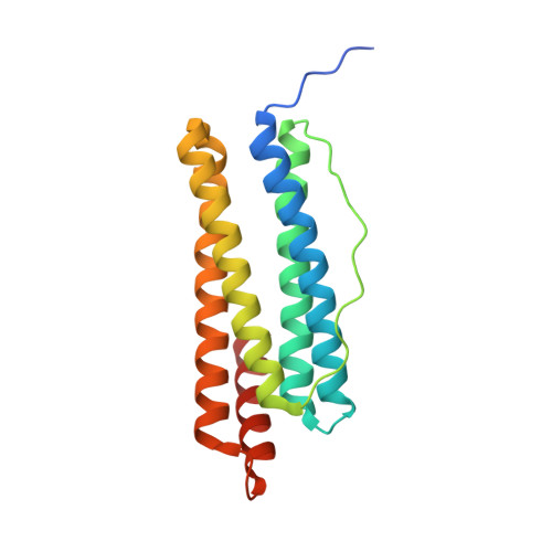
3EGM

Structural basis of iron transport gating in Helicobacter pylori ferritin


Experimental Data Snapshot

  • Method: X-RAY DIFFRACTION
  • Resolution: 2.10 Å
  • R-Value Free: 0.204 
  • R-Value Work: 0.162 
  • R-Value Observed: 0.164 

wwPDB Validation   3D Report Full Report


This is version 1.3 of the entry. See complete history


Literature

The crystal structure of ferritin from Helicobacter pylori reveals unusual conformational changes for iron uptake.

Cho, K.J.Shin, H.J.Lee, J.H.Kim, K.J.Park, S.S.Lee, Y.Lee, C.Park, S.S.Kim, K.H.

(2009) J Mol Biol 390: 83-98

  • DOI: https://doi.org/10.1016/j.jmb.2009.04.078
  • Primary Citation of Related Structures:  
    3EGM

  • PubMed Abstract: 

    The crystal structure of recombinant ferritin from Helicobacter pylori has been determined in its apo, low-iron-bound, intermediate, and high-iron-bound states. Similar to other members of the ferritin family, the bacterial ferritin assembles as a spherical protein shell of 24 subunits, each of which folds into a four-alpha-helix bundle. Significant conformational changes were observed at the BC loop and the entrance of the 4-fold symmetry channel in the intermediate and high-iron-bound states, whereas no change was found in the apo and low-iron-bound states. The imidazole rings of His149 at the channel entrance undergo conformational changes that bear resemblance to heme configuration and are directly coupled to axial translocation of Fe ions through the 4-fold channel. Our results provide the first structural evidence of the translocation of Fe ions through the 4-fold channel in prokaryotes and the transition from a protein-dominated process to a mineral-surface-dominated process during biomineralization.


  • Organizational Affiliation

    Department of Life Sciences and Biotechnology, Korea University, Seoul, Korea.


Macromolecules
Find similar proteins by:  (by identity cutoff)  |  3D Structure
Entity ID: 1
MoleculeChains Sequence LengthOrganismDetailsImage
Ferritin
A, B, C, D, E
A, B, C, D, E, F
181Helicobacter pylori J99Mutation(s): 0 
Gene Names: ftnApfrjhp_0598
EC: 1.16.3.1
UniProt
Find proteins for Q9ZLI1 (Helicobacter pylori (strain J99 / ATCC 700824))
Explore Q9ZLI1 
Go to UniProtKB:  Q9ZLI1
Entity Groups  
Sequence Clusters30% Identity50% Identity70% Identity90% Identity95% Identity100% Identity
UniProt GroupQ9ZLI1
Sequence Annotations
Expand
  • Reference Sequence
Small Molecules
Ligands 2 Unique
IDChains Name / Formula / InChI Key2D Diagram3D Interactions
GOL
Query on GOL

Download Ideal Coordinates CCD File 
J [auth A],
M [auth B],
R [auth D]
GLYCEROL
C3 H8 O3
PEDCQBHIVMGVHV-UHFFFAOYSA-N
FE
Query on FE

Download Ideal Coordinates CCD File 
G [auth A]
H [auth A]
I [auth A]
K [auth B]
L [auth B]
G [auth A],
H [auth A],
I [auth A],
K [auth B],
L [auth B],
N [auth C],
O [auth C],
P [auth D],
Q [auth D],
S [auth E],
T [auth E],
U [auth E],
V [auth F],
W [auth F],
X [auth F]
FE (III) ION
Fe
VTLYFUHAOXGGBS-UHFFFAOYSA-N
Experimental Data & Validation

Experimental Data

  • Method: X-RAY DIFFRACTION
  • Resolution: 2.10 Å
  • R-Value Free: 0.204 
  • R-Value Work: 0.162 
  • R-Value Observed: 0.164 
  • Space Group: I 4
Unit Cell:
Length ( Å )Angle ( ˚ )
a = 128.528α = 90
b = 128.528β = 90
c = 165.38γ = 90
Software Package:
Software NamePurpose
DENZOdata reduction
SCALEPACKdata scaling
REFMACrefinement
PDB_EXTRACTdata extraction
HKL-2000data collection
HKL-2000data reduction
HKL-2000data scaling
MOLREPphasing

Structure Validation

View Full Validation Report


Entry History 

Deposition Data

Revision History  (Full details and data files)

  • Version 1.0: 2009-07-28
    Type: Initial release
  • Version 1.1: 2011-07-13
    Changes: Source and taxonomy, Version format compliance
  • Version 1.2: 2017-10-25
    Changes: Refinement description
  • Version 1.3: 2024-03-20
    Changes: Data collection, Database references, Derived calculations