3EFP | pdb_00003efp

Crystal structure of the Escherichia coli twin arginine leader peptide binding protein DmsD in a monomeric form


Experimental Data Snapshot

  • Method: X-RAY DIFFRACTION
  • Resolution: 2.01 Å
  • R-Value Free: 
    0.213 (Depositor), 0.212 (DCC) 
  • R-Value Work: 
    0.178 (Depositor), 0.179 (DCC) 
  • R-Value Observed: 
    0.180 (Depositor) 

Starting Model: experimental
View more details

wwPDB Validation 3D Report Full Report

Validation slider image for 3EFP

This is version 1.2 of the entry. See complete history

Literature

Structural analysis of a monomeric form of the twin-arginine leader peptide binding chaperone Escherichia coli DmsD.

Stevens, C.M.Winstone, T.M.Turner, R.J.Paetzel, M.

(2009) J Mol Biology 389: 124-133

  • DOI: https://doi.org/10.1016/j.jmb.2009.03.069
  • Primary Citation Related Structures: 
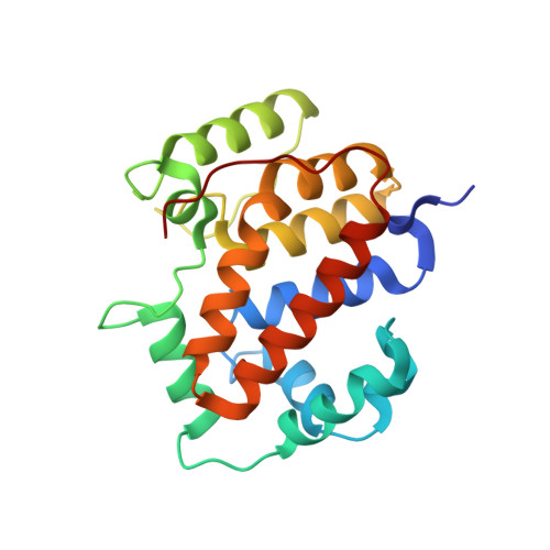
    3EFP

  • PubMed Abstract: 

    The redox enzyme maturation proteins play an essential role in the proofreading and membrane targeting of protein substrates to the twin-arginine translocase. Functionally, the most thoroughly characterized redox enzyme maturation protein to date is Escherichia coli DmsD (EcDmsD). Herein, we present the X-ray crystal structure of the monomeric form of the EcDmsD refined to 2.0 A resolution, with clear electron density present for each of its 204 amino acid residues. The structural data presented here complement the biochemical data previously generated regarding the function of these twin-arginine translocase leader peptide binding chaperone proteins. Docking and molecular dynamics simulation experiments were used to provide a proposed model for how this chaperone is able to recognize the leader peptide of its substrate DmsA. The interactions observed in the model are in agreement with previous biochemical data and suggest intimate interactions between the conserved twin-arginine motif of the leader peptide of E. coli DmsA and the most conserved regions on the surface of EcDmsD.


  • Organizational Affiliation
    • Department of Molecular Biology and Biochemistry, Simon Fraser University, Burnaby, British Columbia, Canada.

Macromolecule Content 

  • Total Structure Weight: 48.98 kDa 
  • Atom Count: 3,860 
  • Modeled Residue Count: 412 
  • Deposited Residue Count: 416 
  • Unique protein chains: 1

Macromolecules

Find similar proteins by:|  3D Structure
Entity ID: 1
MoleculeChains  Sequence LengthOrganismDetailsImage
Twin-arginine leader-binding protein dmsD
A, B
208Escherichia coli K-12Mutation(s): 0 
Gene Names: dmsDynfIb1591JW5262
UniProt
Find proteins for P69853 (Escherichia coli (strain K12))
Explore P69853 
Go to UniProtKB:  P69853
Entity Groups
Sequence Clusters30% Identity50% Identity70% Identity90% Identity95% Identity100% Identity
UniProt GroupP69853
Sequence Annotations
Expand
Reference Sequence

Small Molecules

Ligands 4 Unique
IDChains Name / Formula / InChI Key2D Diagram3D Interactions
TRS

Query on TRS


Download:Ideal Coordinates CCD File
C [auth A],
E [auth A],
M [auth B],
N [auth B]
2-AMINO-2-HYDROXYMETHYL-PROPANE-1,3-DIOL
C4 H12 N O3
LENZDBCJOHFCAS-UHFFFAOYSA-O
GOL

Query on GOL


Download:Ideal Coordinates CCD File
D [auth A]
F [auth A]
G [auth A]
H [auth A]
L [auth B]
D [auth A],
F [auth A],
G [auth A],
H [auth A],
L [auth B],
O [auth B]
GLYCEROL
C3 H8 O3
PEDCQBHIVMGVHV-UHFFFAOYSA-N
CL

Query on CL


Download:Ideal Coordinates CCD File
I [auth A]
J [auth A]
K [auth A]
P [auth B]
Q [auth B]
I [auth A],
J [auth A],
K [auth A],
P [auth B],
Q [auth B],
R [auth B]
CHLORIDE ION
Cl
VEXZGXHMUGYJMC-UHFFFAOYSA-M
NA

Query on NA


Download:Ideal Coordinates CCD File
S [auth B]SODIUM ION
Na
FKNQFGJONOIPTF-UHFFFAOYSA-N

Experimental Data & Validation

Experimental Data

  • Method: X-RAY DIFFRACTION
  • Resolution: 2.01 Å
  • R-Value Free:  0.213 (Depositor), 0.212 (DCC) 
  • R-Value Work:  0.178 (Depositor), 0.179 (DCC) 
  • R-Value Observed: 0.180 (Depositor) 
Space Group: P 31 2 1
Unit Cell:
Length ( Å )Angle ( ˚ )
a = 128.02α = 90
b = 128.02β = 90
c = 78.723γ = 120
Software Package:
Software NamePurpose
d*TREKdata scaling
REFMACrefinement
PDB_EXTRACTdata extraction
CrystalCleardata collection
d*TREKdata reduction
PHASERphasing

Structure Validation

View Full Validation Report


Entry History 

Deposition Data

Revision History  (Full details and data files)

  • Version 1.0: 2009-04-07
    Type: Initial release
  • Version 1.1: 2011-07-13
    Changes: Advisory, Source and taxonomy, Version format compliance
  • Version 1.2: 2023-08-30
    Changes: Data collection, Database references, Derived calculations, Refinement description