3EDL | pdb_00003edl

Kinesin13-Microtubule Ring complex


Experimental Data Snapshot

  • Method: ELECTRON MICROSCOPY
  • Resolution: 28.0 Å
  • Aggregation State: FILAMENT 
  • Reconstruction Method: HELICAL 

wwPDB Validation 3D Report Full Report

Validation slider image for 3EDL

This is version 1.4 of the entry. See complete history

Literature

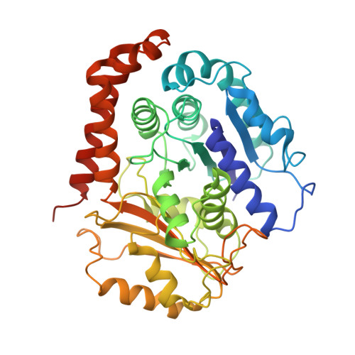
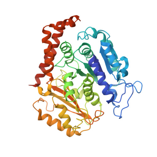
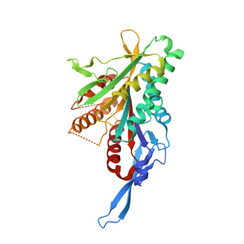
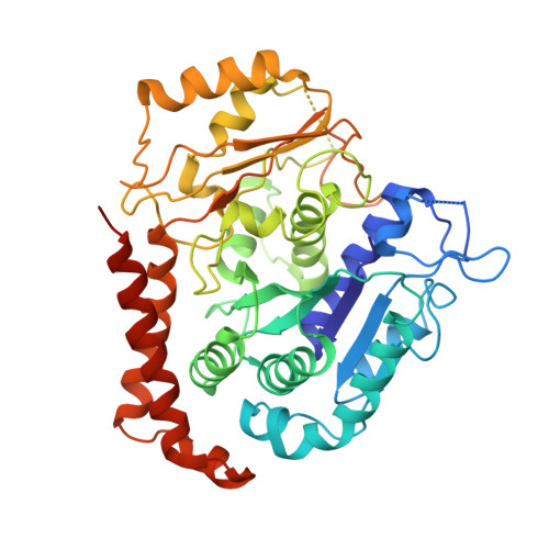
Structure of the kinesin13-microtubule ring complex.

Tan, D.Rice, W.J.Sosa, H.

(2008) Structure 16: 1732-1739

  • DOI: https://doi.org/10.1016/j.str.2008.08.017
  • Primary Citation Related Structures: 
    3EDL

  • PubMed Abstract: 

    To investigate the mechanism of kinesin13-induced microtubule depolymerization, we have calculated a three-dimensional (3D) map of the kinesin13-microtubule ring complex, using cryo-electron microscopy (cryo-EM) and image analysis. An atomic model of the complex was produced by docking the crystal structures of tubulin and a kinesin13 motor domain (MD) into the 3D map. The model reveals a snapshot of the depolymerization mechanism by providing a 3D view of the complex formed between the kinesin13 MD and a curved tubulin protofilament (pf). It suggests that contacts mediated by kinesin13 class-specific residues in the putative microtubule-binding site stabilize intra-dimer tubulin curvature. In addition, a tubulin-binding site on the kinesin13 MD was identified. Mutations at this class-conserved site selectively disrupt the formation of microtubule-associated ring complexes.


  • Organizational Affiliation
    • Department of Physiology and Biophysics, Albert Einstein College of Medicine, Bronx NY, 10461, USA.

Macromolecule Content 

  • Total Structure Weight: 241.02 kDa 
  • Atom Count: 15,757 
  • Modeled Residue Count: 1,979 
  • Deposited Residue Count: 2,123 
  • Unique protein chains: 4

Macromolecules

Find similar proteins by:|  3D Structure
Entity ID: 1
MoleculeChains  Sequence LengthOrganismDetailsImage
Tubulin alpha-1A chain451Bos taurusMutation(s): 0 
EC: 3.6.5
UniProt
Find proteins for P81947 (Bos taurus)
Explore P81947 
Go to UniProtKB:  P81947
Entity Groups
Sequence Clusters30% Identity50% Identity70% Identity90% Identity95% Identity100% Identity
UniProt GroupP81947
Sequence Annotations
Expand
Reference Sequence
Find similar proteins by:|  3D Structure
Entity ID: 2
MoleculeChains  Sequence LengthOrganismDetailsImage
Beta tubulinB,
E [auth G]
445Bos taurusMutation(s): 0 
UniProt
Find proteins for P02554 (Sus scrofa)
Explore P02554 
Go to UniProtKB:  P02554
Entity Groups
Sequence Clusters30% Identity50% Identity70% Identity90% Identity95% Identity100% Identity
UniProt GroupP02554
Sequence Annotations
Expand
Reference Sequence
Find similar proteins by:|  3D Structure
Entity ID: 3
MoleculeChains  Sequence LengthOrganismDetailsImage
Kinesin-like protein KIF2CC [auth D]331Drosophila melanogasterMutation(s): 0 
Gene Names: DmKlp10A
UniProt & NIH Common Fund Data Resources
Find proteins for Q922S8 (Mus musculus)
Explore Q922S8 
Go to UniProtKB:  Q922S8
IMPC:  MGI:1921054
Entity Groups
Sequence Clusters30% Identity50% Identity70% Identity90% Identity95% Identity100% Identity
UniProt GroupQ922S8
Sequence Annotations
Expand
Reference Sequence
Find similar proteins by:|  3D Structure
Entity ID: 4
MoleculeChains  Sequence LengthOrganismDetailsImage
Tubulin alpha-1A chainD [auth F]451Bos taurusMutation(s): 0 
EC: 3.6.5
UniProt
Find proteins for P81947 (Bos taurus)
Explore P81947 
Go to UniProtKB:  P81947
Entity Groups
Sequence Clusters30% Identity50% Identity70% Identity90% Identity95% Identity100% Identity
UniProt GroupP81947
Sequence Annotations
Expand
Reference Sequence

Small Molecules

Ligands 7 Unique
IDChains Name / Formula / InChI Key2D Diagram3D Interactions
TA1

Query on TA1


Download:Ideal Coordinates CCD File
J [auth B]TAXOL
C47 H51 N O14
RCINICONZNJXQF-MZXODVADSA-N
GTP

Query on GTP


Download:Ideal Coordinates CCD File
H [auth B],
N [auth F]
GUANOSINE-5'-TRIPHOSPHATE
C10 H16 N5 O14 P3
XKMLYUALXHKNFT-UUOKFMHZSA-N
ANP

Query on ANP


Download:Ideal Coordinates CCD File
L [auth D]PHOSPHOAMINOPHOSPHONIC ACID-ADENYLATE ESTER
C10 H17 N6 O12 P3
PVKSNHVPLWYQGJ-KQYNXXCUSA-N
GDP

Query on GDP


Download:Ideal Coordinates CCD File
I [auth B],
O [auth F]
GUANOSINE-5'-DIPHOSPHATE
C10 H15 N5 O11 P2
QGWNDRXFNXRZMB-UUOKFMHZSA-N
CN2

Query on CN2


Download:Ideal Coordinates CCD File
P [auth G]2-MERCAPTO-N-[1,2,3,10-TETRAMETHOXY-9-OXO-5,6,7,9-TETRAHYDRO-BENZO[A]HEPTALEN-7-YL]ACETAMIDE
C22 H25 N O6 S
TYDIWMTWTXFWSY-HNNXBMFYSA-N
ZN

Query on ZN


Download:Ideal Coordinates CCD File
F [auth A]ZINC ION
Zn
PTFCDOFLOPIGGS-UHFFFAOYSA-N
MG

Query on MG


Download:Ideal Coordinates CCD File
G [auth A],
K [auth D],
M [auth F]
MAGNESIUM ION
Mg
JLVVSXFLKOJNIY-UHFFFAOYSA-N

Experimental Data & Validation

Experimental Data

  • Method: ELECTRON MICROSCOPY
  • Resolution: 28.0 Å
  • Aggregation State: FILAMENT 
  • Reconstruction Method: HELICAL 
EM Software:
TaskSoftware PackageVersion
RECONSTRUCTIONCustom
RECONSTRUCTIONMRC
RECONSTRUCTIONPHOELIX
RECONSTRUCTIONSUPRIM

Structure Validation

View Full Validation Report


Entry History 

Deposition Data

Revision History  (Full details and data files)

  • Version 1.0: 2009-01-20
    Type: Initial release
  • Version 1.1: 2011-07-13
    Changes: Version format compliance
  • Version 1.2: 2018-07-18
    Changes: Author supporting evidence, Data collection
  • Version 1.3: 2020-06-24
    Changes: Database references, Source and taxonomy, Structure summary
  • Version 1.4: 2024-02-21
    Changes: Data collection, Database references, Derived calculations