


Crystal structure of Dihydrodipicolinate Synthase from Rhodopseudomonas palustris at 2.0A resolution
Satyanarayana, L., Eswaramoorthy, S., Burley, S.K., Swaminathan, S.To be published.
Experimental Data Snapshot
wwPDB Validation 3D Report Full Report

Entity ID: 1 | |||||
|---|---|---|---|---|---|
| Molecule | Chains | Sequence Length | Organism | Details | Image |
| Putative dihydrodipicolinate synthetase | 300 | Rhodopseudomonas palustris | Mutation(s): 0 Gene Names: dapA1, RPA1571 EC: 4.2.1.52 | 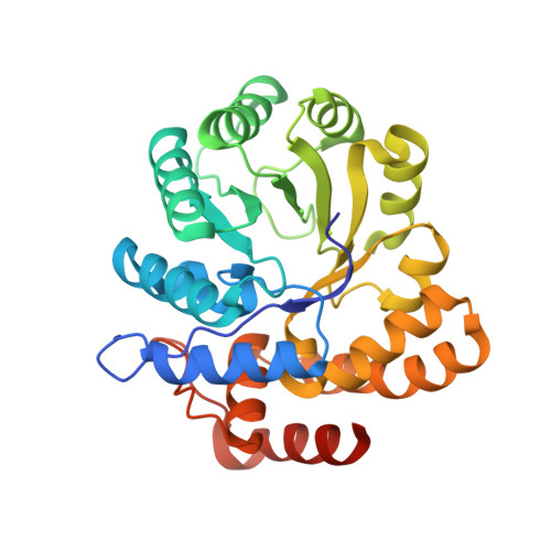 | |
UniProt | |||||
Entity Groups | |||||
| Sequence Clusters | 30% Identity50% Identity70% Identity90% Identity95% Identity100% Identity | ||||
| UniProt Group | Q6N9H8 | ||||
Sequence AnnotationsExpand | |||||
Reference Sequence | |||||
| Ligands 1 Unique | |||||
|---|---|---|---|---|---|
| ID | Chains | Name / Formula / InChI Key | 2D Diagram | 3D Interactions | |
| PGE Download:Ideal Coordinates CCD File | E [auth A], F [auth B] | TRIETHYLENE GLYCOL C6 H14 O4 ZIBGPFATKBEMQZ-UHFFFAOYSA-N |  | ||
| Length ( Å ) | Angle ( ˚ ) |
|---|---|
| a = 79.746 | α = 90 |
| b = 120.852 | β = 90 |
| c = 135.076 | γ = 90 |
| Software Name | Purpose |
|---|---|
| CBASS | data collection |
| SHELXCD | phasing |
| SHARP | phasing |
| CNS | refinement |
| DENZO | data reduction |
| HKL-2000 | data scaling |


